1Z75 | pdb_00001z75

Crystal Structure of ArnA dehydrogenase (decarboxylase) domain, R619M mutant


Experimental Data Snapshot

  • Method: X-RAY DIFFRACTION
  • Resolution: 2.40 Å
  • R-Value Free: 
    0.231 (Depositor), 0.231 (DCC) 
  • R-Value Work: 
    0.203 (Depositor), 0.204 (DCC) 
  • R-Value Observed: 
    0.203 (Depositor) 

wwPDB Validation 3D Report Full Report

Validation slider image for 1Z75

This is version 1.4 of the entry. See complete history

Literature

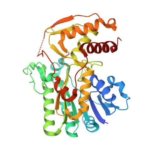
Structure and Mechanism of ArnA: Conformational Change Implies Ordered Dehydrogenase Mechanism in Key Enzyme for Polymyxin Resistance

Gatzeva-Topalova, P.Z.May, A.P.Sousa, M.C.

(2005) Structure 13: 929-942

  • DOI: https://doi.org/10.1016/j.str.2005.03.018
  • Primary Citation Related Structures: 
    1Z73, 1Z74, 1Z75, 1Z7B, 1Z7E

  • PubMed Abstract: 

    The modification of lipid A with 4-amino-4-deoxy-L-arabinose (Ara4N) allows gram-negative bacteria to resist the antimicrobial activity of cationic antimicrobial peptides and antibiotics such as polymyxin. ArnA is the first enzyme specific to the lipid A-Ara4N pathway. It contains two functionally and physically separable domains: a dehydrogenase domain (ArnA_DH) catalyzing the NAD+-dependent oxidative decarboxylation of UDP-Glucuronic acid (UDP-GlcA), and a transformylase domain that formylates UDP-Ara4N. Here, we describe the crystal structure of the full-length bifunctional ArnA with UDP-GlcA and ATP bound to the dehydrogenase domain. Binding of UDP-GlcA triggers a 17 A conformational change in ArnA_DH that opens the NAD+ binding site while trapping UDP-GlcA. We propose an ordered mechanism of substrate binding and product release. Mutation of residues R619 and S433 demonstrates their importance in catalysis and suggests that R619 functions as a general acid in catalysis. The proposed mechanism for ArnA_DH has important implications for the design of selective inhibitors.


  • Organizational Affiliation
    • Department of Chemistry and Biochemistry, University of Colorado at Boulder, Boulder, Colorado 80309, USA.

Macromolecule Content 

  • Total Structure Weight: 41.74 kDa 
  • Atom Count: 2,764 
  • Modeled Residue Count: 334 
  • Deposited Residue Count: 358 
  • Unique protein chains: 1

Macromolecules

Find similar proteins by:|  3D Structure
Entity ID: 1
MoleculeChains  Sequence LengthOrganismDetailsImage
protein ArnA358Escherichia coliMutation(s): 1 
Gene Names: PMRIYFBG
EC: 1.1.1.305 (UniProt), 2.1.2.13 (UniProt)
UniProt
Find proteins for P77398 (Escherichia coli (strain K12))
Explore P77398 
Go to UniProtKB:  P77398
Entity Groups
Sequence Clusters30% Identity50% Identity70% Identity90% Identity95% Identity100% Identity
UniProt GroupP77398
Sequence Annotations
Expand
Reference Sequence

Experimental Data & Validation

Experimental Data

  • Method: X-RAY DIFFRACTION
  • Resolution: 2.40 Å
  • R-Value Free:  0.231 (Depositor), 0.231 (DCC) 
  • R-Value Work:  0.203 (Depositor), 0.204 (DCC) 
  • R-Value Observed: 0.203 (Depositor) 
Space Group: P 41 3 2
Unit Cell:
Length ( Å )Angle ( ˚ )
a = 150.364α = 90
b = 150.364β = 90
c = 150.364γ = 90
Software Package:
Software NamePurpose
DENZOdata reduction
SCALEPACKdata scaling
CNSrefinement
CNSphasing

Structure Validation

View Full Validation Report


Entry History 

Deposition Data

Revision History  (Full details and data files)

  • Version 1.0: 2005-06-07
    Type: Initial release
  • Version 1.1: 2008-04-30
    Changes: Version format compliance
  • Version 1.2: 2011-07-13
    Changes: Derived calculations, Version format compliance
  • Version 1.3: 2021-10-20
    Changes: Database references, Derived calculations
  • Version 1.4: 2024-02-14
    Changes: Data collection