1Z4K

Structure of the D41N variant of the human mitochondrial deoxyribonucleotidase in complex with thymidine 3'-monophosphate


Experimental Data Snapshot

  • Method: X-RAY DIFFRACTION
  • Resolution: 1.75 Å
  • R-Value Free: 0.183 
  • R-Value Work: 0.167 
  • R-Value Observed: 0.168 

wwPDB Validation   3D Report Full Report


Ligand Structure Quality Assessment 


This is version 1.3 of the entry. See complete history


Literature

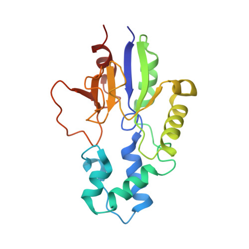
Structural basis for substrate specificity of the human mitochondrial deoxyribonucleotidase

Wallden, K.Ruzzenente, B.Rinaldo-Matthis, A.Bianchi, V.Nordlund, P.

(2005) Structure 13: 1081-1088

  • DOI: https://doi.org/10.1016/j.str.2005.04.023
  • Primary Citation of Related Structures:  
    1Z4I, 1Z4J, 1Z4K, 1Z4L, 1Z4M, 1Z4P, 1Z4Q

  • PubMed Abstract: 

    The human mitochondrial deoxyribonucleotidase catalyzes the dephosphorylation of thymidine and deoxyuridine monophosphates and participates in the regulation of the dTTP pool in mitochondria. We present seven structures of the inactive D41N variant of this enzyme in complex with thymidine 3'-monophosphate, thymidine 5'-monophosphate, deoxyuridine 5'-monophosphate, uridine 5'-monophosphate, deoxyguanosine 5'-monophosphate, uridine 2'-monophosphate, and the 5'-monophosphate of the nucleoside analog 3'-deoxy 2'3'-didehydrothymidine, and we draw conclusions about the substrate specificity based on comparisons with enzyme activities. We show that the enzyme's specificity for the deoxyribo form of nucleoside 5'-monophosphates is due to Ile-133, Phe-49, and Phe-102, which surround the 2' position of the sugar and cause an energetically unfavorable environment for the 2'-hydroxyl group of ribonucleoside 5'-monophosphates. The close binding of the 3'-hydroxyl group of nucleoside 5'-monophosphates to the enzyme indicates that nucleoside analog drugs that are substituted with a bulky group at this position will not be good substrates for this enzyme.


  • Organizational Affiliation

    Department of Biochemistry and Biophysics, Stockholm University, SE-106 91 Stockholm, Sweden.


Macromolecules
Find similar proteins by:  (by identity cutoff)  |  3D Structure
Entity ID: 1
MoleculeChains Sequence LengthOrganismDetailsImage
5'(3')-deoxyribonucleotidase197Homo sapiensMutation(s): 1 
EC: 3.1.3
UniProt & NIH Common Fund Data Resources
Find proteins for Q9NPB1 (Homo sapiens)
Explore Q9NPB1 
Go to UniProtKB:  Q9NPB1
PHAROS:  Q9NPB1
GTEx:  ENSG00000205309 
Entity Groups  
Sequence Clusters30% Identity50% Identity70% Identity90% Identity95% Identity100% Identity
UniProt GroupQ9NPB1
Sequence Annotations
Expand
  • Reference Sequence
Small Molecules
Ligands 2 Unique
IDChains Name / Formula / InChI Key2D Diagram3D Interactions
T3P
Query on T3P

Download Ideal Coordinates CCD File 
C [auth A]THYMIDINE-3'-PHOSPHATE
C10 H15 N2 O8 P
XXYIANZGUOSQHY-XLPZGREQSA-N
MG
Query on MG

Download Ideal Coordinates CCD File 
B [auth A]MAGNESIUM ION
Mg
JLVVSXFLKOJNIY-UHFFFAOYSA-N
Experimental Data & Validation

Experimental Data

  • Method: X-RAY DIFFRACTION
  • Resolution: 1.75 Å
  • R-Value Free: 0.183 
  • R-Value Work: 0.167 
  • R-Value Observed: 0.168 
  • Space Group: P 43 21 2
Unit Cell:
Length ( Å )Angle ( ˚ )
a = 73.18α = 90
b = 73.18β = 90
c = 105.92γ = 90
Software Package:
Software NamePurpose
REFMACrefinement
MOSFLMdata reduction
XDSdata scaling
CNSphasing

Structure Validation

View Full Validation Report


Ligand Structure Quality Assessment 


Entry History 

Deposition Data

Revision History  (Full details and data files)

  • Version 1.0: 2005-07-26
    Type: Initial release
  • Version 1.1: 2008-04-30
    Changes: Version format compliance
  • Version 1.2: 2011-07-13
    Changes: Version format compliance
  • Version 1.3: 2021-11-10
    Changes: Database references, Derived calculations