1Z02

2-Oxoquinoline 8-Monooxygenase Component: Active site Modulation by Rieske-[2fe-2S] Center Oxidation/Reduction


Experimental Data Snapshot

  • Method: X-RAY DIFFRACTION
  • Resolution: 1.80 Å
  • R-Value Free: 0.240 
  • R-Value Work: 0.200 
  • R-Value Observed: 0.200 

wwPDB Validation   3D Report Full Report


This is version 1.5 of the entry. See complete history


Literature

2-Oxoquinoline 8-Monooxygenase Oxygenase Component: Active Site Modulation by Rieske-[2Fe-2S] Center Oxidation/Reduction

Martins, B.M.Svetlitchnaia, T.Dobbek, H.

(2005) Structure 13: 817-824

  • DOI: https://doi.org/10.1016/j.str.2005.03.008
  • Primary Citation of Related Structures:  
    1Z01, 1Z02, 1Z03

  • PubMed Abstract: 

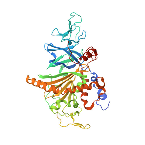
    2-Oxoquinoline 8-monooxygenase is a Rieske non-heme iron oxygenase that catalyzes the NADH-dependent oxidation of the N-heterocyclic aromatic compound 2-oxoquinoline to 8-hydroxy-2-oxoquinoline in the soil bacterium Pseudomonas putida 86. The crystal structure of the oxygenase component of 2-oxoquinoline 8-monooxygenase shows a ring-shaped, C3-symmetric arrangement in which the mononuclear Fe(II) ion active site of one monomer is at a distance of 13 A from the Rieske-[2Fe-2S] center of a second monomer. Structural analyses of oxidized, reduced, and substrate bound states reveal the molecular bases for a new function of Fe-S clusters. Reduction of the Rieske center modulates the mononuclear Fe through a chain of conformational changes across the subunit interface, resulting in the displacement of Fe and its histidine ligand away from the substrate binding site. This creates an additional coordination site at the mononuclear Fe(II) ion and can open a pathway for dioxygen to bind in the substrate-containing active site.


  • Organizational Affiliation

    Laboratorium Proteinkristallographie, Universität Bayreuth, Germany.


Macromolecules
Find similar proteins by:  (by identity cutoff)  |  3D Structure
Entity ID: 1
MoleculeChains Sequence LengthOrganismDetailsImage
2-oxo-1,2-dihydroquinoline 8-monooxygenase, oxygenase component
A, B, C, D, E
A, B, C, D, E, F
446Pseudomonas putidaMutation(s): 0 
EC: 1.14.13.61
UniProt
Find proteins for O05935 (Pseudomonas putida)
Explore O05935 
Go to UniProtKB:  O05935
Entity Groups  
Sequence Clusters30% Identity50% Identity70% Identity90% Identity95% Identity100% Identity
UniProt GroupO05935
Sequence Annotations
Expand
  • Reference Sequence
Small Molecules
Ligands 2 Unique
IDChains Name / Formula / InChI Key2D Diagram3D Interactions
FES
Query on FES

Download Ideal Coordinates CCD File 
H [auth A]
J [auth B]
L [auth C]
N [auth D]
P [auth E]
H [auth A],
J [auth B],
L [auth C],
N [auth D],
P [auth E],
R [auth F]
FE2/S2 (INORGANIC) CLUSTER
Fe2 S2
NIXDOXVAJZFRNF-UHFFFAOYSA-N
FE
Query on FE

Download Ideal Coordinates CCD File 
G [auth A]
I [auth B]
K [auth C]
M [auth D]
O [auth E]
G [auth A],
I [auth B],
K [auth C],
M [auth D],
O [auth E],
Q [auth F]
FE (III) ION
Fe
VTLYFUHAOXGGBS-UHFFFAOYSA-N
Experimental Data & Validation

Experimental Data

  • Method: X-RAY DIFFRACTION
  • Resolution: 1.80 Å
  • R-Value Free: 0.240 
  • R-Value Work: 0.200 
  • R-Value Observed: 0.200 
  • Space Group: P 21 21 21
Unit Cell:
Length ( Å )Angle ( ˚ )
a = 102.03α = 90
b = 169.97β = 90
c = 175.35γ = 90
Software Package:
Software NamePurpose
MAR345data collection
XDSdata reduction
CNSrefinement
XDSdata scaling
CNSphasing

Structure Validation

View Full Validation Report


Entry History 

Deposition Data

Revision History  (Full details and data files)

  • Version 1.0: 2005-05-24
    Type: Initial release
  • Version 1.1: 2008-04-30
    Changes: Version format compliance
  • Version 1.2: 2011-07-13
    Changes: Version format compliance
  • Version 1.3: 2017-10-11
    Changes: Refinement description
  • Version 1.4: 2018-07-18
    Changes: Data collection
  • Version 1.5: 2024-03-13
    Changes: Data collection, Database references, Derived calculations