Structural, mutagenic, and kinetic analysis of the binding of substrates and inhibitors of human phenylethanolamine N-methyltransferase
Wu, Q., Gee, C.L., Lin, F., Tyndall, J.D., Martin, J.L., Grunewald, G.L., McLeish, M.J.(2005) J Med Chem 48: 7243-7252
- PubMed: 16279783 Search on PubMed
- DOI: https://doi.org/10.1021/jm050568o
- Primary Citation Related Structures:
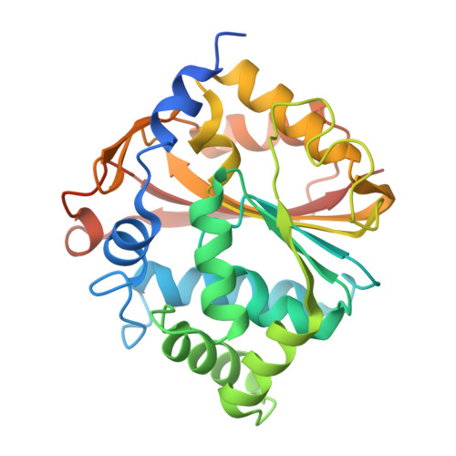
1YZ3 - PubMed Abstract:
The X-ray structure of human phenylethanolamine N-methyltransferase (hPNMT) complexed with its product, S-adenosyl-L-homocysteine (4), and the most potent inhibitor reported to date, SK&F 64139 (7), was used to identify the residues involved in inhibitor binding. Four of these residues, Val53, Lys57, Glu219 and Asp267, were replaced, in turn, with alanine. All variants had increased Km values for phenylethanolamine (10), but only D267A showed a noteworthy (20-fold) decrease in its kcat value. Both WT hPNMT and D267A had similar kcat values for a rigid analogue, anti-9-amino-6-(trifluoromethyl)benzonorbornene (12), suggesting that Asp267 plays an important role in positioning the substrate but does not participate directly in catalysis. The Ki values for the binding of inhibitors such as 7 to the E219A and D267A variants increased by 2-3 orders of magnitude. Further, the inhibitors were shown to bind up to 50-fold more tightly in the presence of S-adenosyl-L-methionine (3), suggesting that the binding of the latter brings about a conformational change in the enzyme.
- Department of Medicinal Chemistry, University of Michigan, 428 Church Street, Ann Arbor, Michigan 48109, USA.
Organizational Affiliation: