1YY8 | pdb_00001yy8

Crystal structure of the Fab fragment from the monoclonal antibody cetuximab/Erbitux/IMC-C225


Experimental Data Snapshot

  • Method: X-RAY DIFFRACTION
  • Resolution: 2.00 Å
  • R-Value Free: 
    0.264 (Depositor), 0.264 (DCC) 
  • R-Value Work: 
    0.224 (Depositor), 0.223 (DCC) 

Starting Models: experimental
View more details

wwPDB Validation 3D Report Full Report

Validation slider image for 1YY8

This is version 1.4 of the entry. See complete history

Literature

Structural basis for inhibition of the epidermal growth factor receptor by cetuximab

Li, S.Schmitz, K.R.Jeffrey, P.D.Wiltzius, J.J.W.Kussie, P.Ferguson, K.M.

(2005) Cancer Cell 7: 301-311

  • DOI: https://doi.org/10.1016/j.ccr.2005.03.003
  • Primary Citation Related Structures: 
    1YY8, 1YY9

  • PubMed Abstract: 

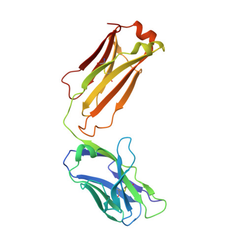
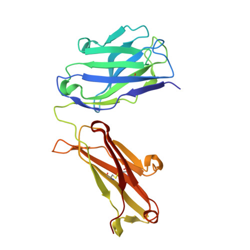
    Recent structural studies of epidermal growth factor receptor (EGFR) family extracellular regions have identified an unexpected mechanism for ligand-induced receptor dimerization that has important implications for activation and inhibition of these receptors. Here we describe the 2.8 angstroms resolution X-ray crystal structure of the antigen binding (Fab) fragment from cetuximab (Erbitux), an inhibitory anti-EGFR antibody, in complex with the soluble extracellular region of EGFR (sEGFR). The sEGFR is in the characteristic "autoinhibited" or "tethered" inactive configuration. Cetuximab interacts exclusively with domain III of sEGFR, partially occluding the ligand binding region on this domain and sterically preventing the receptor from adopting the extended conformation required for dimerization. We suggest that both these effects contribute to potent inhibition of EGFR activation.


  • Organizational Affiliation
    • Department of Physiology, University of Pennsylvania School of Medicine, Philadelphia, Pennsylvania 19104, USA.

Macromolecule Content 

  • Total Structure Weight: 94.03 kDa 
  • Atom Count: 6,958 
  • Modeled Residue Count: 868 
  • Deposited Residue Count: 868 
  • Unique protein chains: 2

Macromolecules

Find similar proteins by:|  3D Structure
Entity ID: 1
MoleculeChains  Sequence LengthOrganismDetailsImage
Cetuximab Fab Light chain
A, C
213Mus musculusHomo sapiens
This entity is chimeric
Mutation(s): 0 
Find similar proteins by:|  3D Structure
Entity ID: 2
MoleculeChains  Sequence LengthOrganismDetailsImage
Cetuximab Fab Heavy chain
B, D
221Mus musculusHomo sapiens
This entity is chimeric
Mutation(s): 0 

Experimental Data & Validation

Experimental Data

  • Method: X-RAY DIFFRACTION
  • Resolution: 2.00 Å
  • R-Value Free:  0.264 (Depositor), 0.264 (DCC) 
  • R-Value Work:  0.224 (Depositor), 0.223 (DCC) 
Space Group: P 21 21 21
Unit Cell:
Length ( Å )Angle ( ˚ )
a = 64.4α = 90
b = 82β = 90
c = 211.4γ = 90
Software Package:
Software NamePurpose
DENZOdata reduction
SCALEPACKdata scaling
AMoREphasing
CNSrefinement

Structure Validation

View Full Validation Report


Entry History 

Deposition Data

Revision History  (Full details and data files)

  • Version 1.0: 2005-04-26
    Type: Initial release
  • Version 1.1: 2008-04-30
    Changes: Version format compliance
  • Version 1.2: 2011-07-13
    Changes: Version format compliance
  • Version 1.3: 2023-08-23
    Changes: Data collection, Database references, Refinement description
  • Version 1.4: 2024-11-20
    Changes: Structure summary