1YQZ

Structure of Coenzyme A-Disulfide Reductase from Staphylococcus aureus refined at 1.54 Angstrom resolution


Experimental Data Snapshot

  • Method: X-RAY DIFFRACTION
  • Resolution: 1.54 Å
  • R-Value Free: 0.206 
  • R-Value Work: 0.177 
  • R-Value Observed: 0.180 

wwPDB Validation   3D Report Full Report


Ligand Structure Quality Assessment 


This is version 1.2 of the entry. See complete history


Literature

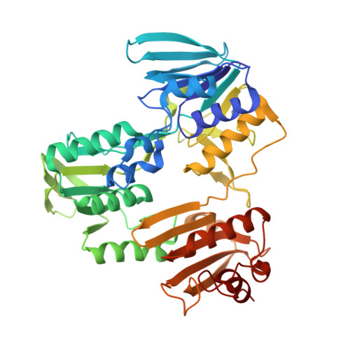
Structure of coenzyme A-disulfide reductase from Staphylococcus aureus at 1.54 A resolution.

Mallett, T.C.Wallen, J.R.Karplus, P.A.Sakai, H.Tsukihara, T.Claiborne, A.

(2006) Biochemistry 45: 11278-11289

  • DOI: https://doi.org/10.1021/bi061139a
  • Primary Citation of Related Structures:  
    1YQZ

  • PubMed Abstract: 

    Coenzyme A (CoASH) replaces glutathione as the major low molecular weight thiol in Staphylococcus aureus; it is maintained in the reduced state by coenzyme A-disulfide reductase (CoADR), a homodimeric enzyme similar to NADH peroxidase but containing a novel Cys43-SSCoA redox center. The crystal structure of S. aureus CoADR has been solved using multiwavelength anomalous dispersion data and refined at a resolution of 1.54 A. The resulting electron density maps define the Cys43-SSCoA disulfide conformation, with Cys43-S(gamma) located at the flavin si face, 3.2 A from FAD-C4aF, and the CoAS- moiety lying in an extended conformation within a cleft at the dimer interface. A well-ordered chloride ion is positioned adjacent to the Cys43-SSCoA disulfide and receives a hydrogen bond from Tyr361'-OH of the complementary subunit, suggesting a role for Tyr361' as an acid-base catalyst during the reduction of CoAS-disulfide. Tyr419'-OH is located 3.2 A from Tyr361'-OH as well and, based on its conservation in known functional CoADRs, also appears to be important for activity. Identification of residues involved in recognition of the CoAS-disulfide substrate and in formation and stabilization of the Cys43-SSCoA redox center has allowed development of a CoAS-binding motif. Bioinformatics analyses indicate that CoADR enzymes are broadly distributed in both bacterial and archaeal kingdoms, suggesting an even broader significance for the CoASH/CoAS-disulfide redox system in prokaryotic thiol/disulfide homeostasis.


  • Organizational Affiliation

    Center for Structural Biology, Wake Forest University School of Medicine, Winston-Salem, North Carolina 27157, USA.


Macromolecules
Find similar proteins by:  (by identity cutoff)  |  3D Structure
Entity ID: 1
MoleculeChains Sequence LengthOrganismDetailsImage
coenzyme A disulfide reductase
A, B
438Staphylococcus aureusMutation(s): 0 
UniProt
Find proteins for O52582 (Staphylococcus aureus (strain NCTC 8325 / PS 47))
Explore O52582 
Go to UniProtKB:  O52582
Entity Groups  
Sequence Clusters30% Identity50% Identity70% Identity90% Identity95% Identity100% Identity
UniProt GroupO52582
Sequence Annotations
Expand
  • Reference Sequence
Small Molecules
Ligands 4 Unique
IDChains Name / Formula / InChI Key2D Diagram3D Interactions
FAD
Query on FAD

Download Ideal Coordinates CCD File 
I [auth A],
O [auth B]
FLAVIN-ADENINE DINUCLEOTIDE
C27 H33 N9 O15 P2
VWWQXMAJTJZDQX-UYBVJOGSSA-N
COA
Query on COA

Download Ideal Coordinates CCD File 
H [auth A],
N [auth B]
COENZYME A
C21 H36 N7 O16 P3 S
RGJOEKWQDUBAIZ-IBOSZNHHSA-N
CL
Query on CL

Download Ideal Coordinates CCD File 
D [auth A]
E [auth A]
F [auth A]
G [auth A]
L [auth B]
D [auth A],
E [auth A],
F [auth A],
G [auth A],
L [auth B],
M [auth B]
CHLORIDE ION
Cl
VEXZGXHMUGYJMC-UHFFFAOYSA-M
MG
Query on MG

Download Ideal Coordinates CCD File 
C [auth A],
J [auth B],
K [auth B]
MAGNESIUM ION
Mg
JLVVSXFLKOJNIY-UHFFFAOYSA-N
Experimental Data & Validation

Experimental Data

  • Method: X-RAY DIFFRACTION
  • Resolution: 1.54 Å
  • R-Value Free: 0.206 
  • R-Value Work: 0.177 
  • R-Value Observed: 0.180 
  • Space Group: P 1 21 1
Unit Cell:
Length ( Å )Angle ( ˚ )
a = 76.06α = 90
b = 65.2β = 104.8
c = 94.49γ = 90
Software Package:
Software NamePurpose
d*TREKdata scaling
d*TREKdata reduction
SOLVEphasing
CNSrefinement

Structure Validation

View Full Validation Report


Ligand Structure Quality Assessment 


Entry History 

Deposition Data

Revision History  (Full details and data files)

  • Version 1.0: 2006-05-09
    Type: Initial release
  • Version 1.1: 2008-04-30
    Changes: Version format compliance
  • Version 1.2: 2011-07-13
    Changes: Version format compliance