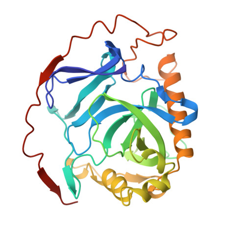
1YPP

ACID ANHYDRIDE HYDROLASE


Experimental Data Snapshot

  • Method: X-RAY DIFFRACTION
  • Resolution: 2.40 Å

wwPDB Validation   3D Report Full Report


This is version 1.4 of the entry. See complete history


Literature

X-ray structure of yeast inorganic pyrophosphatase complexed with manganese and phosphate.

Harutyunyan, E.H.Kuranova, I.P.Vainshtein, B.K.Hohne, W.E.Lamzin, V.S.Dauter, Z.Teplyakov, A.V.Wilson, K.S.

(1996) Eur J Biochem 239: 220-228

  • DOI: https://doi.org/10.1111/j.1432-1033.1996.0220u.x
  • Primary Citation of Related Structures:  
    1YPP

  • PubMed Abstract: 

    The three-dimensional structure of the manganese-phosphate complex of inorganic pyrophosphatase from Saccharomyces cerevisiae has been refined to an R factor of 19.0% at 2.4-A resolution. X-ray data were collected from a single crystal using an imaging plate scanner and synchrotron radiation. There is one dimeric molecule in the asymmetric unit. The upper estimate of the root-mean-square coordinate error is 0.4 A using either the delta A plot or the superposition of the two crystallographically independent subunits. The good agreement between the coordinates of the two subunits, which were not subjected to non-crystallographic symmetry restraints, provides independent validation of the structure analysis. The active site in each subunit contains four manganese ions and two phosphates. The manganese ions are coordinated by the side chains of aspartate and glutamate residues. The phosphate groups, which were identified on the basis of their local stereochemistry, interact either directly or via water molecules with manganese ions and lysine, arginine, and tyrosine side chains. The phosphates are bridged by two of the manganese ions. The outer phosphate is exposed to solvent. The inner phosphate is surrounded by all four manganese ions. The ion-binding sites are related to the order of binding previously established from kinetic studies. A hypothesis for the transition state of the catalytic reaction is put forward.


  • Organizational Affiliation

    Institute of Crystallography, Russian Academy of Sciences, Moscow, Russia.


Macromolecules
Find similar proteins by:  (by identity cutoff)  |  3D Structure
Entity ID: 1
MoleculeChains Sequence LengthOrganismDetailsImage
INORGANIC PYROPHOSPHATASE
A, B
286Saccharomyces cerevisiaeMutation(s): 0 
EC: 3.6.1.1
UniProt
Find proteins for P00817 (Saccharomyces cerevisiae (strain ATCC 204508 / S288c))
Explore P00817 
Go to UniProtKB:  P00817
Entity Groups  
Sequence Clusters30% Identity50% Identity70% Identity90% Identity95% Identity100% Identity
UniProt GroupP00817
Sequence Annotations
Expand
  • Reference Sequence
Small Molecules
Ligands 2 Unique
IDChains Name / Formula / InChI Key2D Diagram3D Interactions
PO4
Query on PO4

Download Ideal Coordinates CCD File 
G [auth A],
H [auth A],
M [auth B],
N [auth B]
PHOSPHATE ION
O4 P
NBIIXXVUZAFLBC-UHFFFAOYSA-K
MN
Query on MN

Download Ideal Coordinates CCD File 
C [auth A]
D [auth A]
E [auth A]
F [auth A]
I [auth B]
C [auth A],
D [auth A],
E [auth A],
F [auth A],
I [auth B],
J [auth B],
K [auth B],
L [auth B]
MANGANESE (II) ION
Mn
WAEMQWOKJMHJLA-UHFFFAOYSA-N
Experimental Data & Validation

Experimental Data

  • Method: X-RAY DIFFRACTION
  • Resolution: 2.40 Å
  • Space Group: P 21 21 21
Unit Cell:
Length ( Å )Angle ( ˚ )
a = 116α = 90
b = 106.2β = 90
c = 56.1γ = 90
Software Package:
Software NamePurpose
PROLSQrefinement
DENZOdata reduction
SCALEPACKdata scaling

Structure Validation

View Full Validation Report


Entry History 

Deposition Data

Revision History  (Full details and data files)

  • Version 1.0: 1996-12-07
    Type: Initial release
  • Version 1.1: 2008-03-24
    Changes: Version format compliance
  • Version 1.2: 2011-07-13
    Changes: Version format compliance
  • Version 1.3: 2017-11-29
    Changes: Derived calculations, Other
  • Version 1.4: 2024-02-14
    Changes: Data collection, Database references, Derived calculations