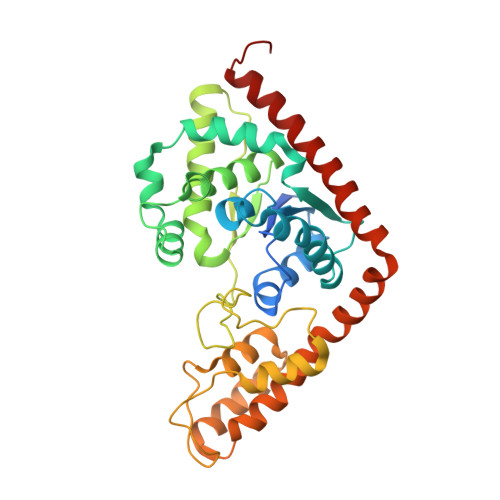
Structures of tryptophanyl-tRNA synthetase II from Deinococcus radiodurans bound to ATP and tryptophan. Insight into subunit cooperativity and domain motions linked to catalysis
Buddha, M.R., Crane, B.R.(2005) J Biological Chem 280: 31965-31973
- PubMed: 15998643
- DOI: https://doi.org/10.1074/jbc.M501568200
- Primary Citation of Related Structures:
1YID, 2A4M - PubMed Abstract:
An auxiliary tryptophanyl tRNA synthetase (drTrpRS II) that interacts with nitric-oxide synthase in the radiation-resistant bacterium Deinococcus radiodurans charges tRNA with tryptophan and 4-nitrotryptophan, a specific nitration product of nitric-oxide synthase. Crystal structures of drTrpRS II, empty of ligands or bound to either Trp or ATP, reveal that drTrpRS II has an overall structure similar to standard bacterial TrpRSs but undergoes smaller amplitude motions of the helical tRNA anti-codon binding (TAB) domain on binding substrates. TAB domain loop conformations that more closely resemble those of human TrpRS than those of Bacillus stearothermophilus TrpRS (bsTrpRS) indicate different modes of tRNA recognition by subclasses of bacterial TrpRSs. A compact state of drTrpRS II binds ATP, from which only minimal TAB domain movement is necessary to bring nucleotide in contact with Trp. However, the signature KMSKS loop of class I synthetases does not completely engage the ATP phosphates, and the adenine ring is not well ordered in the absence of Trp. Thus, progression of the KMSKS loop to a high energy conformation that stages acyl-adenylation requires binding of both substrates. In an asymmetric drTrpRS II dimer, the closed subunit binds ATP, whereas the open subunit binds Trp. A crystallographically symmetric dimer binds no ligands. Half-site reactivity for Trp binding is confirmed by thermodynamic measurements and explained by an asymmetric shift of the dimer interface toward the occupied active site. Upon Trp binding, Asp68 propagates structural changes between subunits by switching its hydrogen bonding partner from dimer interface residue Tyr139 to active site residue Arg30. Since TrpRS IIs are resistant to inhibitors of standard TrpRSs, and pathogens contain drTrpRS II homologs, the structure of drTrpRS II provides a framework for the design of potentially useful antibiotics.
- Department of Chemistry and Chemical Biology, Cornell University, Ithaca, New York 14850, USA.
Organizational Affiliation:


