1Y97 | pdb_00001y97

The human TREX2 3' exonuclease structure suggests a mechanism for efficient non-processive DNA catalysis


Experimental Data Snapshot

  • Method: X-RAY DIFFRACTION
  • Resolution: 2.50 Å
  • R-Value Free: 
    0.261 (Depositor), 0.315 (DCC) 
  • R-Value Work: 
    0.205 (Depositor), 0.265 (DCC) 
  • R-Value Observed: 
    0.205 (Depositor) 

wwPDB Validation 3D Report Full Report

Validation slider image for 1Y97

This is version 1.3 of the entry. See complete history

Literature

The Human TREX2 3' -> 5'-Exonuclease Structure Suggests a Mechanism for Efficient Nonprocessive DNA Catalysis.

Perrino, F.W.Harvey, S.McMillin, S.Hollis, T.

(2005) J Biological Chem 280: 15212-15218

  • DOI: https://doi.org/10.1074/jbc.M500108200
  • Primary Citation Related Structures: 
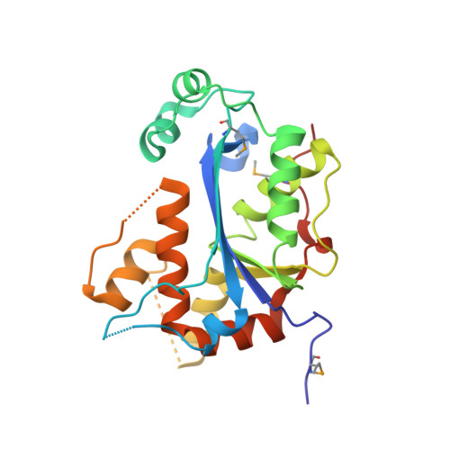
    1Y97

  • PubMed Abstract: 

    The 3' --> 5'-exonucleases process DNA ends in many DNA repair pathways of human cells. Determination of the human TREX2 structure is the first of a dimeric 3'-deoxyribonuclease and indicates how this highly efficient nonprocessive enzyme removes nucleotides at DNA 3' termini. Symmetry in the TREX2 dimer positions the active sites at opposite outer edges providing open access for the DNA. Adjacent to each active site is a flexible region containing three arginines positioned appropriately to bind DNA and to control its entry into the active site. Mutation of these three arginines to alanines reduces the DNA binding capacity by approximately 100-fold with no effect on catalysis. The human TREX2 catalytic residues overlay with the bacterial DnaQ family of 3'-exonucleases confirming the structural conservation of the catalytic sites despite limited sequence identity, and mutations of these residues decrease the still measurable activity by approximately 10(5)-fold, confirming their catalytic role.


  • Organizational Affiliation
    • Department of Biochemistry, Center for Structural Biology, Wake Forest University Health Sciences, Winston-Salem, North Carolina 27157, USA.

Macromolecule Content 

  • Total Structure Weight: 52.45 kDa 
  • Atom Count: 3,340 
  • Modeled Residue Count: 425 
  • Deposited Residue Count: 476 
  • Unique protein chains: 1

Macromolecules

Find similar proteins by:|  3D Structure
Entity ID: 1
MoleculeChains  Sequence LengthOrganismDetailsImage
Three prime repair exonuclease 2
A, B
238Homo sapiensMutation(s): 3 
Gene Names: TREX2
EC: 3.1.11.2
UniProt & NIH Common Fund Data Resources
Find proteins for Q9BQ50 (Homo sapiens)
Explore Q9BQ50 
Go to UniProtKB:  Q9BQ50
PHAROS:  Q9BQ50
GTEx:  ENSG00000183479 
Entity Groups
Sequence Clusters30% Identity50% Identity70% Identity90% Identity95% Identity100% Identity
UniProt GroupQ9BQ50
Sequence Annotations
Expand
Reference Sequence

Small Molecules

Modified Residues  1 Unique
IDChains TypeFormula2D DiagramParent
MSE
Query on MSE
A, B
L-PEPTIDE LINKINGC5 H11 N O2 SeMET

Experimental Data & Validation

Experimental Data

  • Method: X-RAY DIFFRACTION
  • Resolution: 2.50 Å
  • R-Value Free:  0.261 (Depositor), 0.315 (DCC) 
  • R-Value Work:  0.205 (Depositor), 0.265 (DCC) 
  • R-Value Observed: 0.205 (Depositor) 
Space Group: P 21 21 21
Unit Cell:
Length ( Å )Angle ( ˚ )
a = 52.5α = 90
b = 77.2β = 90
c = 101.7γ = 90
Software Package:
Software NamePurpose
HKL-2000data collection
SCALEPACKdata scaling
SOLVEphasing
CNSrefinement
HKL-2000data reduction

Structure Validation

View Full Validation Report


Entry History 

Deposition Data

Revision History  (Full details and data files)

  • Version 1.0: 2005-01-25
    Type: Initial release
  • Version 1.1: 2008-04-30
    Changes: Version format compliance
  • Version 1.2: 2011-07-13
    Changes: Version format compliance
  • Version 1.3: 2024-11-20
    Changes: Data collection, Database references, Derived calculations, Structure summary