1Y4O | pdb_00001y4o

Solution structure of a mouse cytoplasmic Roadblock/LC7 dynein light chain


Experimental Data Snapshot

  • Method: SOLUTION NMR
  • Conformers Calculated: 100 
  • Conformers Submitted: 20 
  • Selection Criteria: target function 

wwPDB Validation 3D Report Full Report

Validation slider image for 1Y4O

This is version 1.4 of the entry. See complete history

Literature

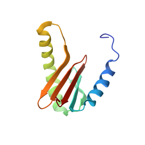
Solution structure of isoform 1 of Roadblock/LC7, a light chain in the dynein complex.

Song, J.Tyler, R.C.Lee, M.S.Tyler, E.M.Markley, J.L.

(2005) J Mol Biology 354: 1043-1051

  • DOI: https://doi.org/10.1016/j.jmb.2005.10.017
  • Primary Citation Related Structures: 
    1Y4O

  • PubMed Abstract: 

    Roadblock/LC7 is a member of a class of dynein light chains involved in regulating the function of the dynein complex. We have determined the three-dimensional structure of isoform 1 of the mouse Roadblock/LC7 cytoplasmic dynein light chain (robl1_mouse) by NMR spectroscopy. In contrast to a previously reported NMR structure of the human homolog with 96% sequence identity (PDB 1TGQ), which showed the protein as a monomer, our results indicate clearly that robl1 exists as a symmetric homodimer. The two beta3-strands pair with each other and form a continuous ten-stranded beta-sheet. The 25-residue alpha2-helix from one subunit packs antiparallel to that of the other subunit on the face of the beta-sheet. Zipper-like hydrophobic contacts between the two helices serve to stabilize the dimer. Through an NMR titration experiment, we localized the site on robl1_mouse that interacts with the 40 residue peptide spanning residues 243 through 282 of IC74-1_rat. These results provide physical evidence for a symmetrical interaction between dimeric robl1 and the two molecules of IC74-1 in the dynein complex.


  • Organizational Affiliation
    • Center for Eukaryotic Structural Genomics, Department of Biochemistry, University of Wisconsin-Madison, WI 53706-1544, USA.

Macromolecule Content 

  • Total Structure Weight: 24 kDa 
  • Atom Count: 1,682 
  • Modeled Residue Count: 208 
  • Deposited Residue Count: 208 
  • Unique protein chains: 1

Macromolecules

Find similar proteins by:|  3D Structure
Entity ID: 1
MoleculeChains  Sequence LengthOrganismDetailsImage
Dynein light chain 2A, cytoplasmic
A, B
104Mus musculusMutation(s): 0 
Gene Names: Dncl2a
UniProt & NIH Common Fund Data Resources
Find proteins for P62627 (Mus musculus)
Explore P62627 
Go to UniProtKB:  P62627
IMPC:  MGI:1914318
Entity Groups
Sequence Clusters30% Identity50% Identity70% Identity90% Identity95% Identity100% Identity
UniProt GroupP62627
Sequence Annotations
Expand
Reference Sequence

Experimental Data & Validation

Experimental Data

  • Method: SOLUTION NMR
  • Conformers Calculated: 100 
  • Conformers Submitted: 20 
  • Selection Criteria: target function 

Structure Validation

View Full Validation Report


Entry History 

Deposition Data

Revision History  (Full details and data files)

  • Version 1.0: 2005-01-18
    Type: Initial release
  • Version 1.1: 2008-04-30
    Changes: Version format compliance
  • Version 1.2: 2011-07-13
    Changes: Version format compliance
  • Version 1.3: 2022-03-02
    Changes: Database references, Derived calculations
  • Version 1.4: 2024-05-22
    Changes: Data collection