1Y47

Structural studies of designed alpha-helical hairpins


Experimental Data Snapshot

  • Method: X-RAY DIFFRACTION
  • Resolution: 2.70 Å
  • R-Value Free: 0.259 
  • R-Value Work: 0.234 
  • R-Value Observed: 0.234 

wwPDB Validation   3D Report Full Report


This is version 1.3 of the entry. See complete history


Literature

Analysis and design of turns in alpha-helical hairpins

Lahr, S.J.Engel, D.E.Stayrook, S.E.Maglio, O.North, B.Geremia, S.Lombardi, A.DeGrado, W.F.

(2005) J Mol Biol 346: 1441-1454

  • DOI: https://doi.org/10.1016/j.jmb.2004.12.016
  • Primary Citation of Related Structures:  
    1MFT, 1U7J, 1U7M, 1Y47

  • PubMed Abstract: 

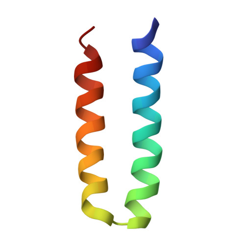
    Although the analysis and design of turns that connect the strands in antiparallel beta-hairpins has reached an advanced state, much less is known concerning turns between antiparallel helices in helical hairpins. We have conducted an analysis of the structures and sequence preferences of two types of interhelical turns, each of which connects the two helices by a two-residue linker in an alphaL-beta conformation. Based on this analysis, it became apparent that the turn introduced into a designed four-helix bundle protein, DF1, did not occur within an optimal structural context. DF1 is a dimeric model for the diiron class of proteins. A longer loop with a beta-alphaR-beta conformation was inserted between two helices in the protein, and a sequence was chosen to stabilize its conformation. X-ray crystallography and NMR analysis of the protein showed the structure to be in excellent agreement with design.


  • Organizational Affiliation

    Department of Biochemistry and Biophysics, University of Pennsylvania, Philadelphia, PA 19104, USA.


Macromolecules
Find similar proteins by:  (by identity cutoff)  |  3D Structure
Entity ID: 1
MoleculeChains Sequence LengthOrganismDetailsImage
dueferri (DF2)
A, B
46N/AMutation(s): 0 
Entity Groups  
Sequence Clusters30% Identity50% Identity70% Identity90% Identity95% Identity100% Identity
Sequence Annotations
Expand
  • Reference Sequence
Experimental Data & Validation

Experimental Data

  • Method: X-RAY DIFFRACTION
  • Resolution: 2.70 Å
  • R-Value Free: 0.259 
  • R-Value Work: 0.234 
  • R-Value Observed: 0.234 
  • Space Group: P 21 21 2
Unit Cell:
Length ( Å )Angle ( ˚ )
a = 48.274α = 90
b = 39.017β = 90
c = 46.891γ = 90
Software Package:
Software NamePurpose
CNSrefinement
DENZOdata reduction
SCALEPACKdata scaling
AMoREphasing

Structure Validation

View Full Validation Report


Entry History 

Deposition Data

Revision History  (Full details and data files)

  • Version 1.0: 2005-03-22
    Type: Initial release
  • Version 1.1: 2008-04-30
    Changes: Version format compliance
  • Version 1.2: 2011-07-13
    Changes: Derived calculations, Version format compliance
  • Version 1.3: 2023-08-23
    Changes: Data collection, Database references, Derived calculations, Refinement description