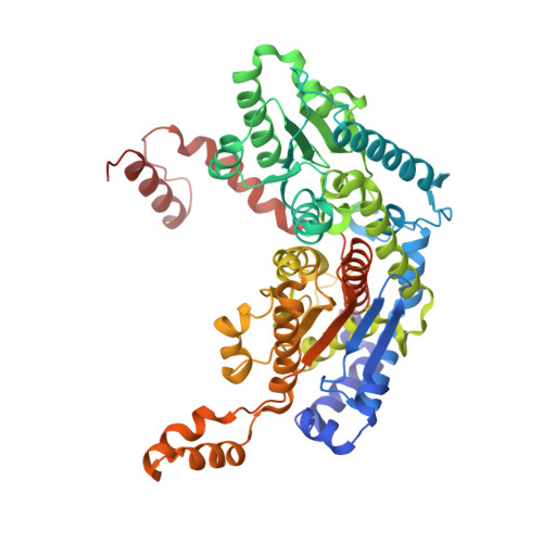
The crystal structure of rabbit phosphoglucose isomerase complexed with D-sorbitol-6-phosphate, an analog of the open chain form of D-glucose-6-phosphate.
Lee, J.H., Jeffery, C.J.(2005) Protein Sci 14: 727-734
- PubMed: 15689508 Search on PubMedSearch on PubMed Central
- DOI: https://doi.org/10.1110/ps.041070205
- Primary Citation Related Structures:
1XTB - PubMed Abstract:
Phosphoglucose isomerase (PGI) catalyzes the isomerization of D-glucose-6-phosphate (G6P) and D-fructose-6-phosphate (F6P) in glycolysis and gluconeogenesis. Analysis of previously reported X-ray crystal structures of PGI without ligand, with the cyclic form of F6P, or with inhibitors that mimic the cis-enediol intermediate led to proposed mechanisms for the ring opening and isomerization steps in the multistep catalytic mechanism. To help complete our model of the overall mechanism, information is needed about the state of PGI between the ring opening and isomerization steps, in other words, a structure of the enzyme complexed with the open form of a substrate or an analog. Here, we report the crystal structure of rabbit PGI complexed with D-sorbitol-6-phosphate (S6P), an analog of the open chain form of G6P, at 2.0 A resolution. As was seen in the PGI/F6P structure, a helix containing amino acid residues 512-520 is found in the "out" position, which provides sufficient space in the active site for a substrate in its cyclic form and which is probably the location of that helix just after ring opening (or just before ring closure). However, the S6P ligand is in an extended conformation, as was seen previously with ligands that mimic the cis-enediol intermediate. The extended conformation enables the ligand to interact with Glu357, which transfers a proton during the isomerization step. The PGI/S6P structure represents the conformation of the enzyme and substrate between the ring opening (or ring closing) step and the isomerization step and helps to complete the model for PGI's catalytic mechanism.
- Laboratory for Molecular Biology, Department of Biological Sciences, MC567, 900 Ashland Ave., University of Illinois at Chicago, Chicago, IL 60607, USA.
Organizational Affiliation: