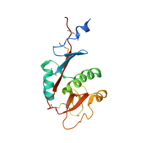
1XPH

Structure of DC-SIGNR and a portion of repeat domain 8


Experimental Data Snapshot

  • Method: X-RAY DIFFRACTION
  • Resolution: 1.41 Å
  • R-Value Free: 0.193 
  • R-Value Work: 0.176 
  • R-Value Observed: 0.177 

wwPDB Validation   3D Report Full Report


This is version 1.3 of the entry. See complete history


Literature

The structure of DC-SIGNR with a portion of its repeat domain lends insights to modeling of the receptor tetramer.

Snyder, G.A.Colonna, M.Sun, P.D.

(2005) J Mol Biol 347: 979-989

  • DOI: https://doi.org/10.1016/j.jmb.2005.01.063
  • Primary Citation of Related Structures:  
    1XPH

  • PubMed Abstract: 

    The dendritic cell-specific ICAM-3 non-integrin (DC-SIGN) and its close relative DC-SIGNR recognize various glycoproteins, both pathogenic and cellular, through the receptor lectin domain-mediated carbohydrate recognition. While the carbohydrate-recognition domains (CRD) exist as monomers and bind individual carbohydrates with low affinity and are permissive in nature, the full-length receptors form tetramers through their repeat domain and recognize specific ligands with high affinity. To understand the tetramer-based ligand binding avidity, we determined the crystal structure of DC-SIGNR with its last repeat region. Compared to the carbohydrate-bound CRD structure, the structure revealed conformational changes in the calcium and carbohydrate coordination loops of CRD, an additional disulfide bond between the N and the C termini of the CRD, and a helical conformation for the last repeat. On the basis of the current crystal structure and other published structures with sequence homology to the repeat domain, we generated a tetramer model for DC-SIGN/R using homology modeling and propose a ligand-recognition index to identify potential receptor ligands.


  • Organizational Affiliation

    Laboratory of Immunogenetics, Structural Immunology Section, National Institute of Allergy and Infectious Diseases, National Institutes of Health, Twinbrook II, 12441 Parklawn Drive, Rockville, MD 20852, USA.


Macromolecules
Find similar proteins by:  (by identity cutoff)  |  3D Structure
Entity ID: 1
MoleculeChains Sequence LengthOrganismDetailsImage
CD209 antigen-like protein 1150Homo sapiensMutation(s): 0 
UniProt & NIH Common Fund Data Resources
Find proteins for Q9H2X3 (Homo sapiens)
Explore Q9H2X3 
Go to UniProtKB:  Q9H2X3
PHAROS:  Q9H2X3
GTEx:  ENSG00000104938 
Entity Groups  
Sequence Clusters30% Identity50% Identity70% Identity90% Identity95% Identity100% Identity
UniProt GroupQ9H2X3
Sequence Annotations
Expand
  • Reference Sequence
Small Molecules
Ligands 1 Unique
IDChains Name / Formula / InChI Key2D Diagram3D Interactions
CA
Query on CA

Download Ideal Coordinates CCD File 
B [auth A]CALCIUM ION
Ca
BHPQYMZQTOCNFJ-UHFFFAOYSA-N
Experimental Data & Validation

Experimental Data

  • Method: X-RAY DIFFRACTION
  • Resolution: 1.41 Å
  • R-Value Free: 0.193 
  • R-Value Work: 0.176 
  • R-Value Observed: 0.177 
  • Space Group: P 21 21 21
Unit Cell:
Length ( Å )Angle ( ˚ )
a = 38.233α = 90
b = 54.887β = 90
c = 62.352γ = 90
Software Package:
Software NamePurpose
REFMACrefinement
HKL-2000data reduction
SCALEPACKdata scaling
AMoREphasing

Structure Validation

View Full Validation Report


Entry History 

Deposition Data

Revision History  (Full details and data files)

  • Version 1.0: 2005-04-19
    Type: Initial release
  • Version 1.1: 2008-04-30
    Changes: Version format compliance
  • Version 1.2: 2011-07-13
    Changes: Version format compliance
  • Version 1.3: 2023-08-23
    Changes: Data collection, Database references, Derived calculations, Refinement description