Conformational Diversity in NAD(H) and Transhydrogenase Nicotinamide Nucleotide Binding Domains
Sundaresan, V., Chartron, J., Yamaguchi, M., Stout, C.D.(2005) J Mol Biology 346: 617-629
- PubMed: 15670609 Search on PubMed
- DOI: https://doi.org/10.1016/j.jmb.2004.11.070
- Primary Citation Related Structures:
1XLT - PubMed Abstract:
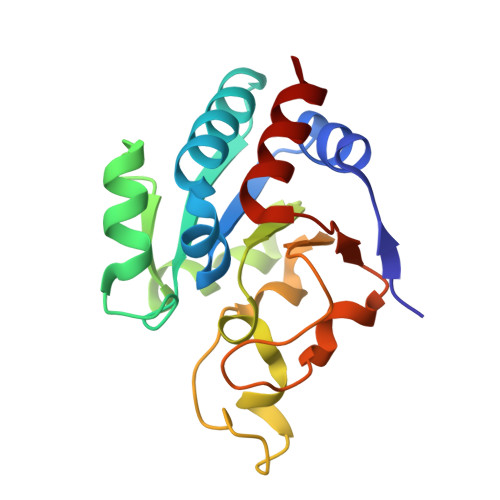
Transhydrogenase (TH) couples direct and stereospecific hydride transfer between NAD(H) and NADP(H), bound within soluble domains I and III, respectively, to proton translocation across membrane bound domain II. The cocrystal structure of Rhodospirillum rubrum TH domains I and III has been determined in the presence of limiting NADH, under conditions in which the subunits reach equilibrium during crystallization. The crystals contain three heterotrimeric complexes, dI(2)dIII, in the asymmetric unit. Multiple conformations of loops and side-chains, and NAD(H) cofactors, are observed in domain I pertaining to substrate/product exchange, and highlighting electrostatic interactions during the hydride transfer. Two interacting NAD(H)-NADPH pairs are observed where alternate conformations of the NAD(H) phosphodiester and conserved arginine side-chains are correlated. In addition, the stereochemistry of one NAD(H)-NADPH pair approaches that expected for nicotinamide hydride transfer reactions. The cocrystal structure exhibits non-crystallographic symmetry that implies another orientation for domain III, which could occur in dimeric TH. Superposition of the "closed" form of domain III (PDB 1PNO, chain A) onto the dI(2)dIII complex reveals a severe steric conflict of highly conserved loops in domains I and III. This overlap, and the overlap with a 2-fold related domain III, suggests that motions of loop D within domain III and of the entire domain are correlated during turnover. The results support the concept that proton pumping in TH is driven by the difference in binding affinity for oxidized and reduced nicotinamide cofactors, and in the absence of a difference in redox potential, must occur through conformational effects.
- Department of Molecular Biology, The Scripps Research Institute, 10550 N. Torrey Pines Road, La Jolla, CA 92037, USA.
Organizational Affiliation: