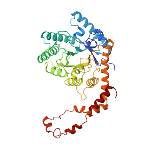
Mechanism for aldose-ketose interconversion by D-xylose isomerase involving ring opening followed by a 1,2-hydride shift.
Collyer, C.A., Henrick, K., Blow, D.M.(1990) J Mol Biology 212: 211-235
- PubMed: 2319597
- DOI: https://doi.org/10.1016/0022-2836(90)90316-E
- Primary Citation of Related Structures:
1XLA, 1XLB, 1XLC, 1XLD, 1XLE, 1XLF, 1XLG, 1XLH, 1XLI, 1XLJ, 1XLK, 1XLL - PubMed Abstract:
The active site and mechanism of D-xylose isomerase have been probed by determination of the crystal structures of the enzyme bound to various substrates, inhibitors and cations. Ring-opening is an obligatory first step of the reaction and is believed to be the rate-determining step for the aldose to ketose conversion. The structure of a complex with a cyclic thio-glucose has been determined and it is concluded that this is an analogue of the Michaelis complex. At -10 degrees C substrates in crystals are observed in the extended chain form. The absence of an appropriately situated base for either the cyclic or extended chain forms from the substrate binding site indicates that the isomerisation does not take place by an enediol or enediolate mechanism. Binding of a trivalent cation places an additional charge at the active site, producing a substrate complex that is analogous to a possible transition state. Of the two binding sites for divalent cations, [1] is permanently occupied under catalytic conditions and is co-ordinated to four carboxylate groups. In the absence of substrate it is exposed to solvent, and in the Michaelis complex analogue, site [1] is octahedrally coordinated, with ligands to O-3 and O-4 of the thiopyranose. In the complex with an open-chain substrate it remains octahedrally co-ordinated, with ligands to O-2 and O-4. Binding at a second cation site [2] is also necessary for catalysis and this site is believed to bind Co2+ more strongly than site [1]. This site is octahedrally co-ordinated to three carboxylate groups (bidentate co-ordination to one of them), an imidazole and a solvent molecule. It is proposed that during the hydride shift the C-O-1 and C-O-2 bonds of the substrate are polarized by the close approach of the site [2] cation. In the transition-state analogue this cation is observed at a site [2'], 1.0 A from site [2] and about 2.7 A from O-1 and O-2 of the substrate. It is likely that co-ordination of the cation to O-1 and O-2 would be concomitant with ionisation of the sugar hydroxyl group. The polarisation of C-O-1 and C-O-2 is assisted by the co-ordination of O-2 to cation [1] and O-1 to a lysine side-chain.(ABSTRACT TRUNCATED AT 400 WORDS)
- Blackett Laboratory, Imperial College, London, England.
Organizational Affiliation:


