1XKF | pdb_00001xkf

Crystal structure of Hypoxic Response Protein I (HRPI) with two coordinated zinc ions


Experimental Data Snapshot

  • Method: X-RAY DIFFRACTION
  • Resolution: 1.90 Å
  • R-Value Free: 
    0.235 (Depositor), 0.234 (DCC) 
  • R-Value Work: 
    0.206 (Depositor), 0.206 (DCC) 

wwPDB Validation 3D Report Full Report

Validation slider image for 1XKF

This is version 1.3 of the entry. See complete history

Literature

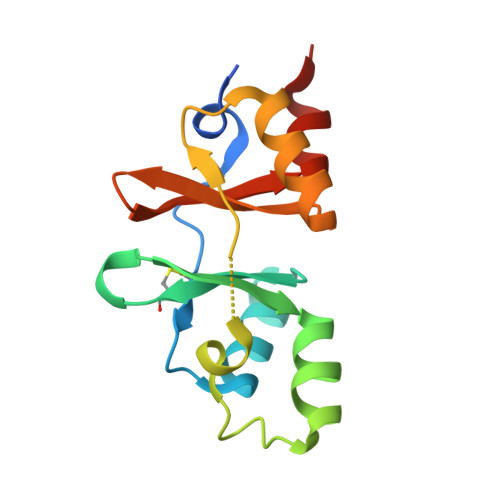
The structure and unusual protein chemistry of hypoxic response protein 1, a latency antigen and highly expressed member of the DosR regulon in Mycobacterium tuberculosis

Sharpe, M.L.Gao, C.Kendall, S.L.Baker, E.N.Lott, J.S.

(2008) J Mol Biology 383: 822-836

  • DOI: https://doi.org/10.1016/j.jmb.2008.07.001
  • Primary Citation Related Structures: 
    1XKF, 1Y5H

  • PubMed Abstract: 

    Mycobacterium tuberculosis adapts to cellular stresses such as decreased oxygen concentration, at least in part, by upregulation of the dormancy survival regulon, which is thought to be important for the bacterium's ability to enter a persistent state in its human host. We have determined the structure of hypoxic response protein 1, a protein encoded by one of the most strongly upregulated genes in the dormancy survival regulon. Hypoxic response protein 1 is an example of a 'cystathionine-beta-synthase-domain-only' protein; however, unlike other cystathionine-beta-synthase domains, it does not appear to bind AMP. The protein is proteolytically sensitive at its C-terminus and contains two unexpected disulfide bonds, one of which appears resistant to reducing agents in solution and is, therefore, most likely buried in the protein and is not solvent-accessible. We show that the protein is secreted from the bacterium in hypoxic in vitro culture and does not accumulate in the bacterial cell wall. The biological function of the protein remains unclear, but we suggest that it may contribute to the modulation of the host immune response. The work reported advances our understanding of the chemistry and cell biology of this intriguing and potentially important protein, and establishes a structural framework for future functional and immunological studies.


  • Organizational Affiliation
    • School of Biological Sciences and Maurice Wilkins Center for Molecular Biodiscovery, University of Auckland, Private Bag 92109, Auckland 1142, New Zealand.

Macromolecule Content 

  • Total Structure Weight: 29.14 kDa 
  • Atom Count: 2,019 
  • Modeled Residue Count: 243 
  • Deposited Residue Count: 266 
  • Unique protein chains: 1

Macromolecules

Find similar proteins by:|  3D Structure
Entity ID: 1
MoleculeChains  Sequence LengthOrganismDetailsImage
hypothetical protein RV2626C
A, B
133Mycobacterium tuberculosisMutation(s): 0 
Gene Names: Rv2626c
UniProt
Find proteins for P9WJA3 (Mycobacterium tuberculosis (strain ATCC 25618 / H37Rv))
Explore P9WJA3 
Go to UniProtKB:  P9WJA3
Entity Groups
Sequence Clusters30% Identity50% Identity70% Identity90% Identity95% Identity100% Identity
UniProt GroupP9WJA3
Sequence Annotations
Expand
Reference Sequence

Experimental Data & Validation

Experimental Data

  • Method: X-RAY DIFFRACTION
  • Resolution: 1.90 Å
  • R-Value Free:  0.235 (Depositor), 0.234 (DCC) 
  • R-Value Work:  0.206 (Depositor), 0.206 (DCC) 
Space Group: P 21 21 21
Unit Cell:
Length ( Å )Angle ( ˚ )
a = 40.65α = 90
b = 50.94β = 90
c = 129.19γ = 90
Software Package:
Software NamePurpose
CNSrefinement
DENZOdata reduction
SCALEPACKdata scaling
AMoREphasing

Structure Validation

View Full Validation Report


Entry History 

Deposition Data

Revision History  (Full details and data files)

  • Version 1.0: 2005-10-11
    Type: Initial release
  • Version 1.1: 2008-01-08
    Changes: Version format compliance
  • Version 1.2: 2011-07-13
    Changes: Version format compliance
  • Version 1.3: 2024-10-23
    Changes: Data collection, Database references, Derived calculations, Structure summary