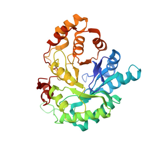
1XF0

Crystal structure of human 17beta-hydroxysteroid dehydrogenase type 5 (AKR1C3) complexed with delta4-androstene-3,17-dione and NADP


Experimental Data Snapshot

  • Method: X-RAY DIFFRACTION
  • Resolution: 2.00 Å
  • R-Value Free: 0.225 
  • R-Value Work: 0.170 
  • R-Value Observed: 0.173 

Starting Model: experimental
View more details

wwPDB Validation   3D Report Full Report


Ligand Structure Quality Assessment 


This is version 1.3 of the entry. See complete history


Literature

Crystal structures of the multispecific 17beta-hydroxysteroid dehydrogenase type 5: critical androgen regulation in human peripheral tissues

Qiu, W.Zhou, M.Labrie, F.Lin, S.-X.

(2004) Mol Endocrinol 18: 1798-1807

  • DOI: https://doi.org/10.1210/me.2004-0032
  • Primary Citation of Related Structures:  
    1XF0

  • PubMed Abstract: 

    Human type 5 17beta-hydroxysteroid dehydrogenase (17beta-HSD5;AKR1C3) plays a major role in the metabolism of androgens in peripheral tissues. In prostate basal cells, this enzyme is involved in the transformation of dehydroepiandrosterone into dihydrotestosterone, the most potent androgen. It is thus a potential target for prostate cancer therapy because it is understood that the testosterone formation by this enzyme is an important factor, particularly in patients who have undergone surgical or medical castration. Here we report the first structure of a human type 5 17beta-HSD in two ternary complexes, in which we found that the androstenedione molecule has a different binding position from that of testosterone. The two testosterone-binding orientations in the substrate-binding site demonstrate the structural basis of the alternative binding and multispecificity of the enzyme. Phe306 and Trp227 are the key residues involved in ligand recognition as well as product release. A safety belt in the cofactor-binding site enhances nicotinamide adenine dinucleotide phosphate binding and accounts for its high affinity as demonstrated by kinetic studies. These structures have provided a dynamic view of the enzyme reaction converting androstenedione to testosterone as well as valuable information for the development of potent enzyme inhibitors.


  • Organizational Affiliation

    Oncology and Molecular Endocrinology Research Center, Centre Hospitalier de l'Université Laval Medical Center (CHUL) and Laval University, Sainte-Foy, Quebec, Canada G1V 4G2.


Macromolecules
Find similar proteins by:  (by identity cutoff)  |  3D Structure
Entity ID: 1
MoleculeChains Sequence LengthOrganismDetailsImage
Aldo-keto reductase family 1 member C3323Homo sapiensMutation(s): 0 
Gene Names: AKR1C3DDH1PGFSKIAA0119
EC: 1.1.1.62 (PDB Primary Data), 1.3.1.20 (PDB Primary Data), 1.1.1.188 (PDB Primary Data)
UniProt & NIH Common Fund Data Resources
Find proteins for P42330 (Homo sapiens)
Explore P42330 
Go to UniProtKB:  P42330
PHAROS:  P42330
GTEx:  ENSG00000196139 
Entity Groups  
Sequence Clusters30% Identity50% Identity70% Identity90% Identity95% Identity100% Identity
UniProt GroupP42330
Sequence Annotations
Expand
  • Reference Sequence
Experimental Data & Validation

Experimental Data

  • Method: X-RAY DIFFRACTION
  • Resolution: 2.00 Å
  • R-Value Free: 0.225 
  • R-Value Work: 0.170 
  • R-Value Observed: 0.173 
  • Space Group: P 63
Unit Cell:
Length ( Å )Angle ( ˚ )
a = 110.579α = 90
b = 110.579β = 90
c = 56.886γ = 120
Software Package:
Software NamePurpose
REFMACrefinement
DENZOdata reduction
SCALEPACKdata scaling
MOLREPphasing

Structure Validation

View Full Validation Report


Ligand Structure Quality Assessment 


Entry History 

Deposition Data

Revision History  (Full details and data files)

  • Version 1.0: 2004-10-26
    Type: Initial release
  • Version 1.1: 2008-04-30
    Changes: Version format compliance
  • Version 1.2: 2011-07-13
    Changes: Version format compliance
  • Version 1.3: 2023-08-23
    Changes: Data collection, Database references, Derived calculations, Refinement description