1XDT | pdb_00001xdt

COMPLEX OF DIPHTHERIA TOXIN AND HEPARIN-BINDING EPIDERMAL GROWTH FACTOR


Experimental Data Snapshot

  • Method: X-RAY DIFFRACTION
  • Resolution: 2.65 Å
  • R-Value Free: 
    0.290 (Depositor) 
  • R-Value Work: 
    0.172 (Depositor), 0.183 (DCC) 
  • R-Value Observed: 
    0.172 (Depositor) 

Starting Model: experimental
View more details

wwPDB Validation 3D Report Full Report

Validation slider image for 1XDT

This is version 1.4 of the entry. See complete history

Literature

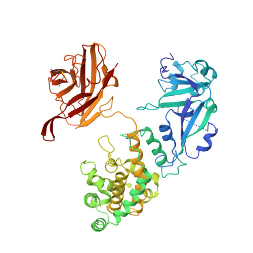
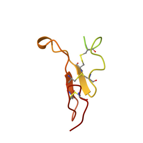
Crystal structure of the complex of diphtheria toxin with an extracellular fragment of its receptor.

Louie, G.V.Yang, W.Bowman, M.E.Choe, S.

(1997) Mol Cell 1: 67-78

  • DOI: https://doi.org/10.1016/s1097-2765(00)80008-8
  • Primary Citation Related Structures: 
    1XDT

  • PubMed Abstract: 

    We describe the crystal structure at 2.65 A resolution of diphtheria toxin (DT) complexed 1:1 with a fragment of its cell-surface receptor, the precursor of heparin-binding epidermal-growth-factor-like growth factor (HBEGF). HBEGF in the complex has the typical EGF-like fold and packs its principal beta hairpin against the face of a beta sheet in the receptor-binding domain of DT. The interface has a predominantly hydrophobic core, and polar interactions are formed at the periphery. The structure of the complex suggests that part of the membrane anchor of the receptor can interact with a hinge region of DT. The toxin molecule is thereby induced to form an open conformation conducive to membrane insertion. The structure provides a basis for altering the binding specificity of the toxin, and may also serve as a model for other EGF-receptor interactions.


  • Organizational Affiliation
    • Structural Biology Laboratory, Salk Institute for Biological Studies, La Jolla, California 92037, USA.

Macromolecule Content 

  • Total Structure Weight: 67.34 kDa 
  • Atom Count: 4,354 
  • Modeled Residue Count: 559 
  • Deposited Residue Count: 614 
  • Unique protein chains: 2

Macromolecules

Find similar proteins by:|  3D Structure
Entity ID: 1
MoleculeChains  Sequence LengthOrganismDetailsImage
DIPHTHERIA TOXINA [auth T]535Corynebacterium diphtheriaeMutation(s): 0 
EC: 2.4.2.36
UniProt
Find proteins for P00588 (Corynephage beta)
Explore P00588 
Go to UniProtKB:  P00588
Entity Groups
Sequence Clusters30% Identity50% Identity70% Identity90% Identity95% Identity100% Identity
UniProt GroupP00588
Sequence Annotations
Expand
Reference Sequence
Find similar proteins by:|  3D Structure
Entity ID: 2
MoleculeChains  Sequence LengthOrganismDetailsImage
HEPARIN-BINDING EPIDERMAL GROWTH FACTORB [auth R]79Homo sapiensMutation(s): 0 
UniProt & NIH Common Fund Data Resources
Find proteins for Q99075 (Homo sapiens)
Explore Q99075 
Go to UniProtKB:  Q99075
PHAROS:  Q99075
GTEx:  ENSG00000113070 
Entity Groups
Sequence Clusters30% Identity50% Identity70% Identity90% Identity95% Identity100% Identity
UniProt GroupQ99075
Sequence Annotations
Expand
Reference Sequence

Experimental Data & Validation

Experimental Data

  • Method: X-RAY DIFFRACTION
  • Resolution: 2.65 Å
  • R-Value Free:  0.290 (Depositor) 
  • R-Value Work:  0.172 (Depositor), 0.183 (DCC) 
  • R-Value Observed: 0.172 (Depositor) 
Space Group: C 2 2 21
Unit Cell:
Length ( Å )Angle ( ˚ )
a = 89.4α = 90
b = 103.7β = 90
c = 127.7γ = 90
Software Package:
Software NamePurpose
DENZOdata reduction
SCALEPACKdata scaling
AMoREphasing
X-PLORrefinement

Structure Validation

View Full Validation Report


Entry History 

Deposition Data

Revision History  (Full details and data files)

  • Version 1.0: 1998-02-25
    Type: Initial release
  • Version 1.1: 2008-03-24
    Changes: Version format compliance
  • Version 1.2: 2011-07-13
    Changes: Derived calculations, Version format compliance
  • Version 1.3: 2023-08-09
    Changes: Database references, Refinement description
  • Version 1.4: 2024-10-23
    Changes: Data collection, Structure summary