1XDF | pdb_00001xdf

Crystal structure of pathogenesis-related protein LlPR-10.2A from yellow lupine


Experimental Data Snapshot

  • Method: X-RAY DIFFRACTION
  • Resolution: 1.90 Å
  • R-Value Free: 
    0.251 (Depositor), 0.257 (DCC) 
  • R-Value Work: 
    0.205 (Depositor), 0.213 (DCC) 
  • R-Value Observed: 
    0.207 (Depositor) 

Starting Model: experimental
View more details

wwPDB Validation 3D Report Full Report

Validation slider image for 1XDF

This is version 1.3 of the entry. See complete history

Literature

Structure of a yellow lupin pathogenesis-related PR-10 protein belonging to a novel subclass.

Pasternak, O.Biesiadka, J.Dolot, R.Handschuh, L.Bujacz, G.Sikorski, M.M.Jaskolski, M.

(2005) Acta Crystallogr D Biol Crystallogr 61: 99-107

  • DOI: https://doi.org/10.1107/S0907444904028173
  • Primary Citation Related Structures: 
    1XDF

  • PubMed Abstract: 

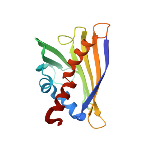
    Pathogenesis-related (PR) proteins of class 10 are abundant in higher plants. Some of these proteins are induced under stress conditions as part of the plant defence mechanism. Other homologues are developmentally regulated and their expression varies in different plant organs. The PR-10 proteins are encoded by multigene families, have a weight of about 17 kDa and are found in the cytosol. In yellow lupin, nine different homologues have been identified and divided into two subclasses, LlPR-10.1 and LlPR-10.2. Within each subclass the sequence identity is about 75-91%, while across the subclasses it is only 59-60%. Here, the crystal structure of a yellow lupin PR-10 protein from the second subclass, LlPR-10.2A, is presented. The structure was solved by molecular replacement and refined to R = 0.205 using 1.9 A resolution data. The general fold of LlPR-10.2A resembles that of the other PR-10 proteins and consists of a long C-terminal alpha-helix surrounded by a seven-stranded antiparallel beta-sheet, with two shorter alpha-helices located between strands beta1 and beta2. The most variable part of the structure, the C-terminal helix, is strongly kinked towards the beta-sheet core in both LlPR-10.2A molecules present in the asymmetric unit. This unexpected feature reduces the size of the hydrophobic cavity observed in other PR-10 proteins that is reported to be the ligand-binding site. As in other PR-10 structures, a surface loop located near the entrance to the cavity shows very high structural conservation and stability despite the high glycine content in its sequence.


  • Organizational Affiliation
    • Institute of Bioorganic Chemistry, Polish Academy of Sciences, Poznan, Poland.

Macromolecule Content 

  • Total Structure Weight: 33.85 kDa 
  • Atom Count: 2,643 
  • Modeled Residue Count: 314 
  • Deposited Residue Count: 314 
  • Unique protein chains: 1

Macromolecules

Find similar proteins by:|  3D Structure
Entity ID: 1
MoleculeChains  Sequence LengthOrganismDetailsImage
PR10.2A
A, B
157Lupinus luteusMutation(s): 0 
Gene Names: Llpr-10.2a
EC: 3.1.27
UniProt
Find proteins for Q9LLQ3 (Lupinus luteus)
Explore Q9LLQ3 
Go to UniProtKB:  Q9LLQ3
Entity Groups
Sequence Clusters30% Identity50% Identity70% Identity90% Identity95% Identity100% Identity
UniProt GroupQ9LLQ3
Sequence Annotations
Expand
Reference Sequence

Small Molecules

Ligands 2 Unique
IDChains Name / Formula / InChI Key2D Diagram3D Interactions
EPE

Query on EPE


Download:Ideal Coordinates CCD File
D [auth A]4-(2-HYDROXYETHYL)-1-PIPERAZINE ETHANESULFONIC ACID
C8 H18 N2 O4 S
JKMHFZQWWAIEOD-UHFFFAOYSA-N
NA

Query on NA


Download:Ideal Coordinates CCD File
C [auth A]SODIUM ION
Na
FKNQFGJONOIPTF-UHFFFAOYSA-N

Experimental Data & Validation

Experimental Data

  • Method: X-RAY DIFFRACTION
  • Resolution: 1.90 Å
  • R-Value Free:  0.251 (Depositor), 0.257 (DCC) 
  • R-Value Work:  0.205 (Depositor), 0.213 (DCC) 
  • R-Value Observed: 0.207 (Depositor) 
Space Group: P 21 21 21
Unit Cell:
Length ( Å )Angle ( ˚ )
a = 48.956α = 90
b = 69.243β = 90
c = 112.918γ = 90
Software Package:
Software NamePurpose
REFMACrefinement
DENZOdata reduction
SCALEPACKdata scaling
AMoREphasing

Structure Validation

View Full Validation Report


Entry History 

Deposition Data

Revision History  (Full details and data files)

  • Version 1.0: 2005-02-15
    Type: Initial release
  • Version 1.1: 2008-04-30
    Changes: Version format compliance
  • Version 1.2: 2011-07-13
    Changes: Advisory, Version format compliance
  • Version 1.3: 2023-10-25
    Changes: Data collection, Database references, Derived calculations, Refinement description