1XCR | pdb_00001xcr

Crystal Structure of Longer Splice Variant of PTD012 from Homo sapiens reveals a novel Zinc-containing fold


Experimental Data Snapshot

  • Method: X-RAY DIFFRACTION
  • Resolution: 1.70 Å
  • R-Value Free: 
    0.222 (Depositor), 0.228 (DCC) 
  • R-Value Work: 
    0.190 (Depositor), 0.198 (DCC) 

Starting Model: other
View more details

wwPDB Validation 3D Report Full Report

Validation slider image for 1XCR

This is version 1.4 of the entry. See complete history

Literature

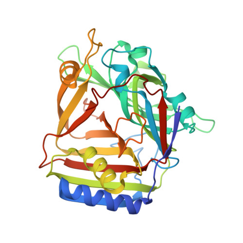
Crystal structure of Homo sapiens PTD012 reveals a zinc-containing hydrolase fold

Manjasetty, B.A.Buessow, K.Fieber-Erdmann, M.Roske, Y.Gobom, J.Scheich, C.Goetz, F.Niesen, F.H.Heinemann, U.

(2006) Protein Sci 15: 914-920

  • DOI: https://doi.org/10.1110/ps.052037006
  • Primary Citation Related Structures: 
    1XCR

  • PubMed Abstract: 

    The human protein PTD012 is the longer product of an alternatively spliced gene and was described to be localized in the nucleus. The X-ray structure analysis at 1.7 A resolution of PTD012 through SAD phasing reveals a monomeric protein and a novel fold. The shorter splice form was also studied and appears to be unfolded and non-functional. The structure of PTD012 displays an alphabetabetaalpha four-layer topology. A metal ion residing between the central beta-sheets is partially coordinated by three histidine residues. X-ray absorption near-edge structure (XANES) analysis identifies the PTD012-bound ion as Zn(2+). Tetrahedral coordination of the ion is completed by the carboxylate oxygen atom of an acetate molecule taken up from the crystallization buffer. The binding of Zn(2+) to PTD012 is reminiscent of zinc-containing enzymes such as carboxypeptidase, carbonic anhydrase, and beta-lactamase. Biochemical assays failed to demonstrate any of these enzyme activities in PTD012. However, PTD012 exhibits ester hydrolase activity on the substrate p-nitrophenyl acetate.


  • Organizational Affiliation
    • Protein Structure Factory, Berlin 12489, Germany.

Macromolecule Content 

  • Total Structure Weight: 70.6 kDa 
  • Atom Count: 5,180 
  • Modeled Residue Count: 626 
  • Deposited Residue Count: 632 
  • Unique protein chains: 1

Macromolecules

Find similar proteins by:|  3D Structure
Entity ID: 1
MoleculeChains  Sequence LengthOrganismDetailsImage
hypothetical protein PTD012
A, B
316Homo sapiensMutation(s): 0 
Gene Names: DKFZp564H1122
EC: 4.1.1.34
UniProt & NIH Common Fund Data Resources
Find proteins for Q9H0W9 (Homo sapiens)
Explore Q9H0W9 
Go to UniProtKB:  Q9H0W9
PHAROS:  Q9H0W9
GTEx:  ENSG00000182919 
Entity Groups
Sequence Clusters30% Identity50% Identity70% Identity90% Identity95% Identity100% Identity
UniProt GroupQ9H0W9
Sequence Annotations
Expand
Reference Sequence

Experimental Data & Validation

Experimental Data

  • Method: X-RAY DIFFRACTION
  • Resolution: 1.70 Å
  • R-Value Free:  0.222 (Depositor), 0.228 (DCC) 
  • R-Value Work:  0.190 (Depositor), 0.198 (DCC) 
Space Group: P 1 21 1
Unit Cell:
Length ( Å )Angle ( ˚ )
a = 44.572α = 90
b = 52.888β = 88.89
c = 124.485γ = 90
Software Package:
Software NamePurpose
REFMACrefinement
DENZOdata reduction
SCALEPACKdata scaling
SOLVEphasing

Structure Validation

View Full Validation Report


Entry History 

Deposition Data

Revision History  (Full details and data files)

  • Version 1.0: 2005-09-27
    Type: Initial release
  • Version 1.1: 2008-04-30
    Changes: Version format compliance
  • Version 1.2: 2011-07-13
    Changes: Advisory, Version format compliance
  • Version 1.3: 2024-03-13
    Changes: Data collection, Database references, Derived calculations
  • Version 1.4: 2024-04-03
    Changes: Refinement description