1XCG | pdb_00001xcg

Crystal Structure of Human RhoA in complex with DH/PH fragment of PDZRHOGEF


Experimental Data Snapshot

  • Method: X-RAY DIFFRACTION
  • Resolution: 2.50 Å
  • R-Value Free: 
    0.281 (Depositor) 
  • R-Value Work: 
    0.224 (Depositor) 
  • R-Value Observed: 
    0.235 (Depositor) 

wwPDB Validation 3D Report Full Report

Validation slider image for 1XCG

This is version 1.4 of the entry. See complete history

Literature

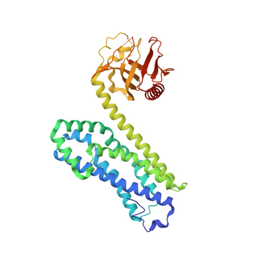
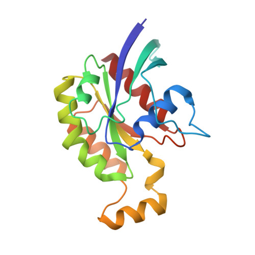
The crystal structure of RhoA in complex with the DH/PH fragment of PDZRhoGEF, an activator of the Ca(2+) sensitization pathway in smooth muscle

Derewenda, U.Oleksy, A.Stevenson, A.S.Korczynska, J.Dauter, Z.Somlyo, A.P.Otlewski, J.Somlyo, A.V.Derewenda, Z.S.

(2004) Structure 12: 1955-1965

  • DOI: https://doi.org/10.1016/j.str.2004.09.003
  • Primary Citation Related Structures: 
    1XCG

  • PubMed Abstract: 

    Calcium sensitization in smooth muscle is mediated by the RhoA GTPase, activated by hitherto unspecified nucleotide exchange factors (GEFs) acting downstream of Galphaq/Galpha(12/13) trimeric G proteins. Here, we show that at least one potential GEF, the PDZRhoGEF, is present in smooth muscle, and its isolated DH/PH fragment induces calcium sensitization in the absence of agonist-mediated signaling. In vitro, the fragment shows high selectivity for the RhoA GTPase. Full-length fragment is required for the nucleotide exchange, as the isolated DH domain enhances it only marginally. We crystallized the DH/PH fragment of PDZRhoGEF in complex with nonprenylated human RhoA and determined the structure at 2.5 A resolution. The refined molecular model reveals that the mutual disposition of the DH and PH domains is significantly different from other previously described complexes involving DH/PH tandems, and that the PH domain interacts with RhoA in a unique mode. The DH domain makes several specific interactions with RhoA residues not conserved among other Rho family members, suggesting the molecular basis for the observed specificity.


  • Organizational Affiliation
    • Department of Molecular Physiology and Biological Physics, University of Virginia, Charlottesville, VA 22908, USA.

Macromolecule Content 

  • Total Structure Weight: 125.88 kDa 
  • Atom Count: 8,750 
  • Modeled Residue Count: 1,069 
  • Deposited Residue Count: 1,092 
  • Unique protein chains: 2

Macromolecules

Find similar proteins by:|  3D Structure
Entity ID: 1
MoleculeChains  Sequence LengthOrganismDetailsImage
Rho guanine nucleotide exchange factor 11A,
C [auth E]
368Homo sapiensMutation(s): 0 
Gene Names: ARHGEF11KIAA0380
UniProt & NIH Common Fund Data Resources
Find proteins for O15085 (Homo sapiens)
Explore O15085 
Go to UniProtKB:  O15085
PHAROS:  O15085
GTEx:  ENSG00000132694 
Entity Groups
Sequence Clusters30% Identity50% Identity70% Identity90% Identity95% Identity100% Identity
UniProt GroupO15085
Sequence Annotations
Expand
Reference Sequence
Find similar proteins by:|  3D Structure
Entity ID: 2
MoleculeChains  Sequence LengthOrganismDetailsImage
Transforming protein RhoAB,
D [auth F]
178Homo sapiensMutation(s): 1 
Gene Names: RHOAARHAARH12RHO12
EC: 3.6.5.2
UniProt & NIH Common Fund Data Resources
Find proteins for P61586 (Homo sapiens)
Explore P61586 
Go to UniProtKB:  P61586
PHAROS:  P61586
GTEx:  ENSG00000067560 
Entity Groups
Sequence Clusters30% Identity50% Identity70% Identity90% Identity95% Identity100% Identity
UniProt GroupP61586
Sequence Annotations
Expand
Reference Sequence

Experimental Data & Validation

Experimental Data

  • Method: X-RAY DIFFRACTION
  • Resolution: 2.50 Å
  • R-Value Free:  0.281 (Depositor) 
  • R-Value Work:  0.224 (Depositor) 
  • R-Value Observed: 0.235 (Depositor) 
Space Group: P 1 21 1
Unit Cell:
Length ( Å )Angle ( ˚ )
a = 89.236α = 90
b = 118.782β = 114.34
c = 90.13γ = 90
Software Package:
Software NamePurpose
HKL-2000data collection
SCALEPACKdata scaling
SHELXDphasing
REFMACrefinement
HKL-2000data reduction

Structure Validation

View Full Validation Report


Entry History 

Deposition Data

Revision History  (Full details and data files)

  • Version 1.0: 2004-12-14
    Type: Initial release
  • Version 1.1: 2008-04-30
    Changes: Version format compliance
  • Version 1.2: 2011-07-13
    Changes: Version format compliance
  • Version 1.3: 2021-10-20
    Changes: Database references
  • Version 1.4: 2024-02-14
    Changes: Data collection, Refinement description