1WW5

Agrocybe cylindracea galectin complexed with 3'-sulfonyl lactose


Experimental Data Snapshot

  • Method: X-RAY DIFFRACTION
  • Resolution: 2.20 Å
  • R-Value Free: 0.267 
  • R-Value Work: 0.198 
  • R-Value Observed: 0.198 

wwPDB Validation   3D Report Full Report


This is version 2.1 of the entry. See complete history


Literature

Structural Basis of a Fungal Galectin from Agrocybe cylindracea for Recognizing Sialoconjugate

Ban, M.Yoon, H.J.Demirkan, E.Utsumi, S.Mikami, B.Yagi, F.

(2005) J Mol Biol 351: 695-706

  • DOI: https://doi.org/10.1016/j.jmb.2005.06.045
  • Primary Citation of Related Structures:  
    1WW4, 1WW5, 1WW6, 1WW7

  • PubMed Abstract: 

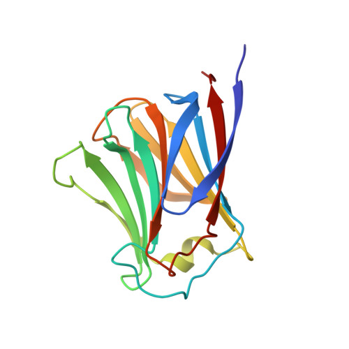
    Galectin from an edible fungus Agrocybe cylindracea (ACG) has a strong preference for N-acetylneuraminyl lactose (NeuAcalpha2-3lactose). The sugar recognition mechanism of ACG was explored by the X-ray crystallographic analyses of ligand-free ACG, and its complex with lactose, 3'-sulfonyl lactose and NeuAcalpha2-3lactose. The refined structure shows that ACG is a "proto"-type galectin composed of a beta-sandwich of two antiparallel sheets, each with six strands, in contrast to the five and six strands in animal galectins. ACG dimer in solution was classified as being among the "layer"-type. The carbohydrate recognition domain (CRD) of this galectin is common to those of animal galectins, except for substitution of one residue, Ala64, which corresponds to Asn46 in human galectin 1. A five-residue insertion in ACG at positions 42-46 involving Ser44 and Asn46 modified the architecture of the sugar binding site that contributes sialic acid specificity. Furthermore, it was found that the binding of a sulfate ion near the CRD in the ligand-free form led to a change in the conformation of the loop region caused by main-chain cis/trans transition between Ser44 and Pro45.


  • Organizational Affiliation

    Laboratory of Food Design and Development, Graduate school of Agriculture, Kyoto University, Kyoto, Japan.


Macromolecules
Find similar proteins by:  (by identity cutoff)  |  3D Structure
Entity ID: 1
MoleculeChains Sequence LengthOrganismDetailsImage
galectin
A, B, C, D
160Agrocybe cylindraceaMutation(s): 0 
UniProt
Find proteins for Q6WY08 (Cyclocybe aegerita)
Explore Q6WY08 
Go to UniProtKB:  Q6WY08
Entity Groups  
Sequence Clusters30% Identity50% Identity70% Identity90% Identity95% Identity100% Identity
UniProt GroupQ6WY08
Sequence Annotations
Expand
  • Reference Sequence
Oligosaccharides

Help

Entity ID: 2
MoleculeChains Length2D Diagram Glycosylation3D Interactions
3-O-sulfo-beta-D-galactopyranose-(1-4)-beta-D-glucopyranose
E, F, G, H
2N/A
Glycosylation Resources
GlyTouCan:  G20044TD
GlyCosmos:  G20044TD
GlyGen:  G20044TD
Experimental Data & Validation

Experimental Data

  • Method: X-RAY DIFFRACTION
  • Resolution: 2.20 Å
  • R-Value Free: 0.267 
  • R-Value Work: 0.198 
  • R-Value Observed: 0.198 
  • Space Group: P 1
Unit Cell:
Length ( Å )Angle ( ˚ )
a = 47.623α = 89.96
b = 57.648β = 111.65
c = 64.643γ = 90.14
Software Package:
Software NamePurpose
CNSrefinement
SADIEdata reduction
SAINTdata reduction
SAINTdata scaling
CNSphasing

Structure Validation

View Full Validation Report


Entry History 

Deposition Data

Revision History  (Full details and data files)

  • Version 1.0: 2005-08-23
    Type: Initial release
  • Version 1.1: 2008-04-30
    Changes: Version format compliance
  • Version 1.2: 2011-07-13
    Changes: Version format compliance
  • Version 2.0: 2020-07-29
    Type: Remediation
    Reason: Carbohydrate remediation
    Changes: Atomic model, Data collection, Derived calculations, Structure summary
  • Version 2.1: 2024-03-13
    Changes: Data collection, Database references, Structure summary