1WUU

crystal structure of human galactokinase complexed with MgAMPPNP and galactose


Experimental Data Snapshot

  • Method: X-RAY DIFFRACTION
  • Resolution: 2.50 Å
  • R-Value Free: 0.258 
  • R-Value Work: 0.201 
  • R-Value Observed: 0.205 

wwPDB Validation   3D Report Full Report


Ligand Structure Quality Assessment 


This is version 1.3 of the entry. See complete history


Literature

Molecular Structure of Human Galactokinase: IMPLICATIONS FOR TYPE II GALACTOSEMIA

Thoden, J.B.Timson, D.J.Reece, R.J.Holden, H.M.

(2005) J Biol Chem 280: 9662-9670

  • DOI: https://doi.org/10.1074/jbc.M412916200
  • Primary Citation of Related Structures:  
    1WUU

  • PubMed Abstract: 

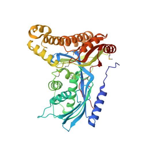
    Galactokinase functions in the Leloir pathway for galactose metabolism by catalyzing the MgATP-dependent phosphorylation of the C-1 hydroxyl group of alpha-D-galactose. The enzyme is known to belong to the GHMP superfamily of small molecule kinases and has attracted significant research attention for well over 40 years. Approximately 20 mutations have now been identified in human galactokinase, which result in the diseased state referred to as Type II galactosemia. Here we report the three-dimensional architecture of human galactokinase with bound alpha-D-galactose and Mg-AMPPNP. The overall fold of the molecule can be described in terms of two domains with the active site wedged between them. The N-terminal domain is dominated by a six-stranded mixed beta-sheet whereas the C-terminal motif contains six alpha-helices and two layers of anti-parallel beta-sheet. Those residues specifically involved in sugar binding include Arg37, Glu43, His44, Asp46, Gly183, Asp186, and Tyr236. The C-1 hydroxyl group of alpha-D-galactose sits within 3.3 A of the gamma-phosphorus of the nucleotide and 3.4 A of the guanidinium group of Arg37. The carboxylate side chain of Asp186 lies within approximately 3.2 A of the C-2 hydroxyl group of alpha-D-galactose and the guanidinium group of Arg37. Both Arg37 and Asp186 are strictly conserved among both prokaryotic and eukaryotic galactokinases. In addition to providing molecular insight into the active site geometry of the enzyme, the model also provides a structural framework upon which to more fully understand the consequences of the those mutations known to give rise to Type II galactosemia.


  • Organizational Affiliation

    Department of Biochemistry, University of Wisconsin, Madison, Wisconsin 53706, USA.


Macromolecules
Find similar proteins by:  (by identity cutoff)  |  3D Structure
Entity ID: 1
MoleculeChains Sequence LengthOrganismDetailsImage
Galactokinase
A, B, C, D
399Homo sapiensMutation(s): 8 
Gene Names: GALK1
EC: 2.7.1.6
UniProt & NIH Common Fund Data Resources
Find proteins for P51570 (Homo sapiens)
Explore P51570 
Go to UniProtKB:  P51570
PHAROS:  P51570
GTEx:  ENSG00000108479 
Entity Groups  
Sequence Clusters30% Identity50% Identity70% Identity90% Identity95% Identity100% Identity
UniProt GroupP51570
Sequence Annotations
Expand
  • Reference Sequence
Small Molecules
Ligands 3 Unique
IDChains Name / Formula / InChI Key2D Diagram3D Interactions
ANP
Query on ANP

Download Ideal Coordinates CCD File 
G [auth A],
J [auth B],
M [auth C],
P [auth D]
PHOSPHOAMINOPHOSPHONIC ACID-ADENYLATE ESTER
C10 H17 N6 O12 P3
PVKSNHVPLWYQGJ-KQYNXXCUSA-N
GLA
Query on GLA

Download Ideal Coordinates CCD File 
E [auth A],
H [auth B],
K [auth C],
N [auth D]
alpha-D-galactopyranose
C6 H12 O6
WQZGKKKJIJFFOK-PHYPRBDBSA-N
MG
Query on MG

Download Ideal Coordinates CCD File 
F [auth A],
I [auth B],
L [auth C],
O [auth D]
MAGNESIUM ION
Mg
JLVVSXFLKOJNIY-UHFFFAOYSA-N
Modified Residues  1 Unique
IDChains TypeFormula2D DiagramParent
MSE
Query on MSE
A, B, C, D
L-PEPTIDE LINKINGC5 H11 N O2 SeMET
Experimental Data & Validation

Experimental Data

  • Method: X-RAY DIFFRACTION
  • Resolution: 2.50 Å
  • R-Value Free: 0.258 
  • R-Value Work: 0.201 
  • R-Value Observed: 0.205 
  • Space Group: P 1 21 1
Unit Cell:
Length ( Å )Angle ( ˚ )
a = 73.2α = 90
b = 109.6β = 95.9
c = 115.8γ = 90
Software Package:
Software NamePurpose
MOSFLMdata reduction
HKL-2000data reduction
SOLVEphasing
TNTrefinement
HKL-2000data scaling

Structure Validation

View Full Validation Report


Ligand Structure Quality Assessment 


Entry History 

Deposition Data

Revision History  (Full details and data files)

  • Version 1.0: 2004-12-28
    Type: Initial release
  • Version 1.1: 2008-04-30
    Changes: Version format compliance
  • Version 1.2: 2011-07-13
    Changes: Version format compliance
  • Version 1.3: 2020-07-29
    Type: Remediation
    Reason: Carbohydrate remediation
    Changes: Advisory, Data collection, Database references, Derived calculations, Structure summary