1WOK | pdb_00001wok

Crystal structure of catalytic domain of human poly(ADP-ribose) polymerase complexed with a quinoxaline-type inhibitor


Experimental Data Snapshot

  • Method: X-RAY DIFFRACTION
  • Resolution: 3.00 Å
  • R-Value Free: 
    0.288 (Depositor), 0.283 (DCC) 
  • R-Value Work: 
    0.233 (Depositor), 0.229 (DCC) 

Starting Model: experimental
View more details

wwPDB Validation 3D Report Full Report

Validation slider image for 1WOK

Ligand Structure Quality Assessment 


This is version 1.4 of the entry. See complete history

Literature

Discovery of quinazolinone and quinoxaline derivatives as potent and selective poly(ADP-ribose) polymerase-1/2 inhibitors.

Iwashita, A.Hattori, K.Yamamoto, H.Ishida, J.Kido, Y.Kamijo, K.Murano, K.Miyake, H.Kinoshita, T.Warizaya, M.Ohkubo, M.Matsuoka, N.Mutoh, S.

(2005) FEBS Lett 579: 1389-1393

  • DOI: https://doi.org/10.1016/j.febslet.2005.01.036
  • Primary Citation Related Structures: 
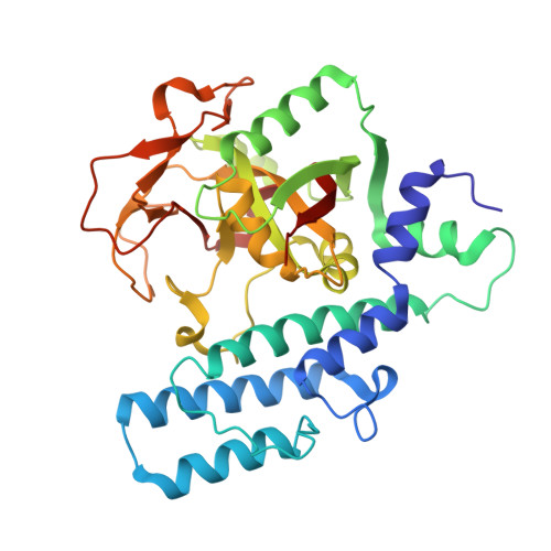
    1WOK

  • PubMed Abstract: 

    Two classes of quinazolinone derivatives and quinoxaline derivatives were identified as potent and selective poly(ADP-ribose) polymerase-1 and 2 (PARP-1) and (PARP-2) inhibitors, respectively. In PARP enzyme assays using recombinant PARP-1 and PARP-2, quinazolinone derivatives displayed relatively high selectivity for PARP-1 and quinoxaline derivatives showed superior selectivity for PARP-2. SBDD analysis via a combination of X-ray structural study and homology modeling suggested distinct interactions of inhibitors with PARP-1 and PARP-2. These findings provide a new structural framework for the design of selective inhibitors for PARP-1 and PARP-2.


  • Organizational Affiliation
    • Medicinal Biology Research Laboratories, Fujisawa Pharmaceutical Co., Ltd., 2-1-6 Kashima, Osaka 532-8514, Japan. aki_iwashita@po.fujisawa.co.jp

Macromolecule Content 

  • Total Structure Weight: 157.92 kDa 
  • Atom Count: 11,096 
  • Modeled Residue Count: 1,400 
  • Deposited Residue Count: 1,400 
  • Unique protein chains: 1

Macromolecules

Find similar proteins by:|  3D Structure
Entity ID: 1
MoleculeChains  Sequence LengthOrganismDetailsImage
Poly [ADP-ribose] polymerase-1
A, B, C, D
350Homo sapiensMutation(s): 0 
Gene Names: PPOL
EC: 2.4.2.30 (PDB Primary Data), 2.4.2 (UniProt)
UniProt & NIH Common Fund Data Resources
Find proteins for P09874 (Homo sapiens)
Explore P09874 
Go to UniProtKB:  P09874
PHAROS:  P09874
GTEx:  ENSG00000143799 
Entity Groups
Sequence Clusters30% Identity50% Identity70% Identity90% Identity95% Identity100% Identity
UniProt GroupP09874
Sequence Annotations
Expand
Reference Sequence

Experimental Data & Validation

Experimental Data

  • Method: X-RAY DIFFRACTION
  • Resolution: 3.00 Å
  • R-Value Free:  0.288 (Depositor), 0.283 (DCC) 
  • R-Value Work:  0.233 (Depositor), 0.229 (DCC) 
Space Group: P 1 21 1
Unit Cell:
Length ( Å )Angle ( ˚ )
a = 90.05α = 90
b = 77.08β = 117.43
c = 113.72γ = 90
Software Package:
Software NamePurpose
CNXrefinement
CCP4data scaling
AMoREphasing

Structure Validation

View Full Validation Report


Ligand Structure Quality Assessment 


Entry History 

Revision History  (Full details and data files)

  • Version 1.0: 2005-03-15
    Type: Initial release
  • Version 1.1: 2008-04-30
    Changes: Version format compliance
  • Version 1.2: 2011-07-13
    Changes: Version format compliance
  • Version 1.3: 2017-10-11
    Changes: Refinement description
  • Version 1.4: 2023-10-25
    Changes: Data collection, Database references, Derived calculations, Refinement description