Structure of the N-terminal Domain of PEX1 AAA-ATPase: CHARACTERIZATION OF A PUTATIVE ADAPTOR-BINDING DOMAIN
Shiozawa, K., Maita, N., Tomii, K., Seto, A., Goda, N., Akiyama, Y., Shimizu, T., Shirakawa, M., Hiroaki, H.(2004) J Biological Chem 279: 50060-50068
- PubMed: 15328346 Search on PubMed
- DOI: https://doi.org/10.1074/jbc.M407837200
- Primary Citation Related Structures:
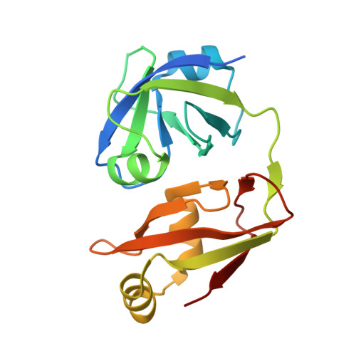
1WLF - PubMed Abstract:
Peroxisomes are responsible for several pathways in primary metabolism, including beta-oxidation and lipid biosynthesis. PEX1 and PEX6 are hexameric AAA-type ATPases, both of which are indispensable in targeting over 50 peroxisomal resident proteins from the cytosol to the peroxisomes. Although the tandem AAA-ATPase domains in the central region of PEX1 and PEX6 are highly similar, the N-terminal sequences are unique. To better understand the distinct molecular function of these two proteins, we analyzed the unique N-terminal domain (NTD) of PEX1. Extensive computational analysis revealed weak similarity (<10% identity) of PEX1 NTD to the N-terminal domains of other membrane-related type II AAA-ATPases, such as VCP (p97) and NSF. We have determined the crystal structure of mouse PEX1 NTD at 2.05-A resolution, which clearly demonstrated that the domain belongs to the double-psi-barrel fold family found in the other AAA-ATPases. The N-domains of both VCP and NSF are structural neighbors of PEX1 NTD with a 2.7- and 2.1-A root mean square deviation of backbone atoms, respectively. Our findings suggest that the supradomain architecture, which is composed of a single N-terminal domain followed by tandem AAA domains, is a common feature of organellar membrane-associating AAA-ATPases. We propose that PEX1 functions as a protein unfoldase in peroxisomal biogenesis, using its N-terminal putative adaptor-binding domain.
- Graduate School of Integrated Science, Yokohama City University, 1-7-29 Suehiro, Tsurumi-ku, Yokohama, Kanagawa 230-0045, Japan.
Organizational Affiliation: