1WHS

STRUCTURE OF THE COMPLEX OF L-BENZYLSUCCINATE WITH WHEAT SERINE CARBOXYPEPTIDASE II AT 2.0 ANGSTROMS RESOLUTION


Experimental Data Snapshot

  • Method: X-RAY DIFFRACTION
  • Resolution: 2.00 Å
  • R-Value Observed: 0.175 

wwPDB Validation   3D Report Full Report


This is version 2.0 of the entry. See complete history


Literature

Structure of the complex of L-benzylsuccinate with wheat serine carboxypeptidase II at 2.0-A resolution.

Bullock, T.L.Branchaud, B.Remington, S.J.

(1994) Biochemistry 33: 11127-11134

  • DOI: https://doi.org/10.1021/bi00203a009
  • Primary Citation of Related Structures:  
    1WHS, 1WHT

  • PubMed Abstract: 

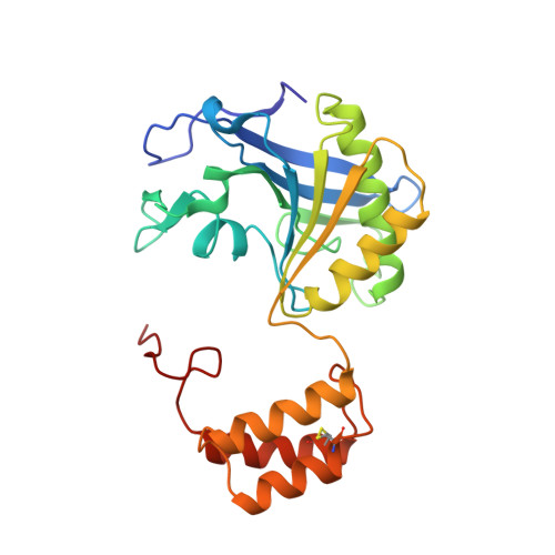
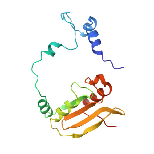
    The structure of the complex of L-benzylsuccinate (Ki = 0.2 mM) bound to wheat serine carboxypeptidase II has been analyzed at 2.0-A resolution for native and inhibited crystals at -170 degrees C. The model has been refined and has a standard crystallographic R-factor of 0.176 for 57,734 reflections observed between 20.0- and 2.0-A resolution. The root mean square deviation from ideal bonds is 0.017 A and from ideal angles is 2.6 degrees. The model consists of 400 amino acids, 4 N-linked saccharide residues, and 430 water molecules. L-Benzylsuccinate occupies a narrow slot in the active site defined by Tyr 60, Tyr 239, and the polypeptide backbone. One carboxylate forms hydrogen bonds to Glu 145, Asn 51, the amide of Gly 52, and the catalytic His 397, suggestive of how the peptide C-terminal carboxylate is recognized by the enzyme. The phenyl ring stacks between Tyr 239 and Tyr 60, while the other carboxylate occupies the "oxyanion hole". One of the oxygens accepts hydrogen bonds from the amides of Tyr 147 and Gly 53, while the other forms a very close contact (2.3 A) with the O gamma of Ser 146, forcing the side chain into a conformation alternative to that found in the resting state of the enzyme. The inhibitor occupies the active site in a way that suggests that it can be regarded as a transition-state analogue of serine carboxypeptidases. The model suggests a novel enzymatic mechanism, involving substrate-assisted catalysis, that might account for the low pH optimum (4.0-5.5) of peptidase activity unique to this family of serine proteinases.


  • Organizational Affiliation

    Institute of Molecular Biology, University of Oregon, Eugene 97403, USA.


Macromolecules
Find similar proteins by:  (by identity cutoff)  |  3D Structure
Entity ID: 1
MoleculeChains Sequence LengthOrganismDetailsImage
SERINE CARBOXYPEPTIDASE II255Triticum aestivumMutation(s): 0 
EC: 3.4.16.1
UniProt
Find proteins for P08819 (Triticum aestivum)
Explore P08819 
Go to UniProtKB:  P08819
Entity Groups  
Sequence Clusters30% Identity50% Identity70% Identity90% Identity95% Identity100% Identity
UniProt GroupP08819
Sequence Annotations
Expand
  • Reference Sequence
Find similar proteins by:  (by identity cutoff)  |  3D Structure
Entity ID: 2
MoleculeChains Sequence LengthOrganismDetailsImage
SERINE CARBOXYPEPTIDASE II153Triticum aestivumMutation(s): 0 
EC: 3.4.16.1
UniProt
Find proteins for P08819 (Triticum aestivum)
Explore P08819 
Go to UniProtKB:  P08819
Entity Groups  
Sequence Clusters30% Identity50% Identity70% Identity90% Identity95% Identity100% Identity
UniProt GroupP08819
Sequence Annotations
Expand
  • Reference Sequence
Oligosaccharides

Help

Entity ID: 3
MoleculeChains Length2D Diagram Glycosylation3D Interactions
alpha-L-fucopyranose-(1-3)-[2-acetamido-2-deoxy-beta-D-glucopyranose-(1-4)]2-acetamido-2-deoxy-beta-D-glucopyranose
C
3N/A
Glycosylation Resources
GlyTouCan:  G11971MR
GlyCosmos:  G11971MR
GlyGen:  G11971MR
Entity ID: 4
MoleculeChains Length2D Diagram Glycosylation3D Interactions
2-acetamido-2-deoxy-beta-D-glucopyranose-(1-4)-2-acetamido-2-deoxy-beta-D-glucopyranose
D
2N/A
Glycosylation Resources
GlyTouCan:  G42666HT
GlyCosmos:  G42666HT
GlyGen:  G42666HT
Small Molecules
Ligands 3 Unique
IDChains Name / Formula / InChI Key2D Diagram3D Interactions
NAG
Query on NAG

Download Ideal Coordinates CCD File 
E [auth A]2-acetamido-2-deoxy-beta-D-glucopyranose
C8 H15 N O6
OVRNDRQMDRJTHS-FMDGEEDCSA-N
GOL
Query on GOL

Download Ideal Coordinates CCD File 
F [auth A]GLYCEROL
C3 H8 O3
PEDCQBHIVMGVHV-UHFFFAOYSA-N
ACY
Query on ACY

Download Ideal Coordinates CCD File 
G [auth A],
H [auth B]
ACETIC ACID
C2 H4 O2
QTBSBXVTEAMEQO-UHFFFAOYSA-N
Experimental Data & Validation

Experimental Data

  • Method: X-RAY DIFFRACTION
  • Resolution: 2.00 Å
  • R-Value Observed: 0.175 
  • Space Group: P 41 21 2
Unit Cell:
Length ( Å )Angle ( ˚ )
a = 95.6α = 90
b = 95.6β = 90
c = 208.8γ = 90
Software Package:
Software NamePurpose
TNTrefinement

Structure Validation

View Full Validation Report


Entry History 

Deposition Data

Revision History  (Full details and data files)

  • Version 1.0: 1994-06-22
    Type: Initial release
  • Version 1.1: 2008-03-03
    Changes: Version format compliance
  • Version 1.2: 2011-07-13
    Changes: Non-polymer description, Version format compliance
  • Version 1.3: 2017-11-29
    Changes: Advisory, Derived calculations, Other
  • Version 2.0: 2020-07-29
    Type: Remediation
    Reason: Carbohydrate remediation
    Changes: Advisory, Atomic model, Data collection, Derived calculations, Structure summary