1WD3

Crystal structure of arabinofuranosidase


Experimental Data Snapshot

  • Method: X-RAY DIFFRACTION
  • Resolution: 1.75 Å
  • R-Value Free: 0.209 
  • R-Value Work: 0.191 
  • R-Value Observed: 0.191 

wwPDB Validation   3D Report Full Report


This is version 2.0 of the entry. See complete history


Literature

Crystal structure of a family 54 alpha-L-arabinofuranosidase reveals a novel carbohydrate-binding module that can bind arabinose

Miyanaga, A.Koseki, T.Matsuzawa, H.Wakagi, T.Shoun, H.Fushinobu, S.

(2004) J Biol Chem 279: 44907-44914

  • DOI: https://doi.org/10.1074/jbc.M405390200
  • Primary Citation of Related Structures:  
    1WD3, 1WD4

  • PubMed Abstract: 

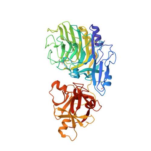
    As the first known structures of a glycoside hydrolase family 54 (GH54) enzyme, we determined the crystal structures of free and arabinose-complex forms of Aspergillus kawachii IFO4308 alpha-l-arabinofuranosidase (AkAbfB). AkAbfB comprises two domains: a catalytic domain and an arabinose-binding domain (ABD). The catalytic domain has a beta-sandwich fold similar to those of clan-B glycoside hydrolases. ABD has a beta-trefoil fold similar to that of carbohydrate-binding module (CBM) family 13. However, ABD shows a number of characteristics distinctive from those of CBM family 13, suggesting that it could be classified into a new CBM family. In the arabinose-complex structure, one of three arabinofuranose molecules is bound to the catalytic domain through many interactions. Interestingly, a disulfide bond formed between two adjacent cysteine residues recognized the arabinofuranose molecule in the active site. From the location of this arabinofuranose and the results of a mutational study, the nucleophile and acid/base residues were determined to be Glu(221) and Asp(297), respectively. The other two arabinofuranose molecules are bound to ABD. The O-1 atoms of the two arabinofuranose molecules bound at ABD are both pointed toward the solvent, indicating that these sites can both accommodate an arabinofuranose side-chain moiety linked to decorated arabinoxylans.


  • Organizational Affiliation

    Department of Biotechnology, The University of Tokyo, 1-1-1 Yayoi, Bunkyo-ku, Tokyo 113-8657, Japan.


Macromolecules
Find similar proteins by:  (by identity cutoff)  |  3D Structure
Entity ID: 1
MoleculeChains Sequence LengthOrganismDetailsImage
alpha-L-arabinofuranosidase B482Aspergillus luchuensisMutation(s): 0 
EC: 3.2.1.55
UniProt
Find proteins for Q8NK89 (Aspergillus kawachii (strain NBRC 4308))
Explore Q8NK89 
Go to UniProtKB:  Q8NK89
Entity Groups  
Sequence Clusters30% Identity50% Identity70% Identity90% Identity95% Identity100% Identity
UniProt GroupQ8NK89
Sequence Annotations
Expand
  • Reference Sequence
Oligosaccharides

Help

Entity ID: 2
MoleculeChains Length2D Diagram Glycosylation3D Interactions
2-acetamido-2-deoxy-beta-D-glucopyranose-(1-4)-2-acetamido-2-deoxy-beta-D-glucopyranose
B
2N-Glycosylation
Glycosylation Resources
GlyTouCan:  G42666HT
GlyCosmos:  G42666HT
GlyGen:  G42666HT
Experimental Data & Validation

Experimental Data

  • Method: X-RAY DIFFRACTION
  • Resolution: 1.75 Å
  • R-Value Free: 0.209 
  • R-Value Work: 0.191 
  • R-Value Observed: 0.191 
  • Space Group: P 21 21 21
Unit Cell:
Length ( Å )Angle ( ˚ )
a = 39.455α = 90
b = 96.894β = 90
c = 145.853γ = 90
Software Package:
Software NamePurpose
CNSrefinement
HKL-2000data reduction
SCALEPACKdata scaling
SOLVEphasing

Structure Validation

View Full Validation Report


Entry History 

Deposition Data

Revision History  (Full details and data files)

  • Version 1.0: 2004-09-14
    Type: Initial release
  • Version 1.1: 2008-04-30
    Changes: Version format compliance
  • Version 1.2: 2011-07-13
    Changes: Non-polymer description, Version format compliance
  • Version 2.0: 2020-07-29
    Type: Remediation
    Reason: Carbohydrate remediation
    Changes: Advisory, Atomic model, Data collection, Database references, Derived calculations, Structure summary