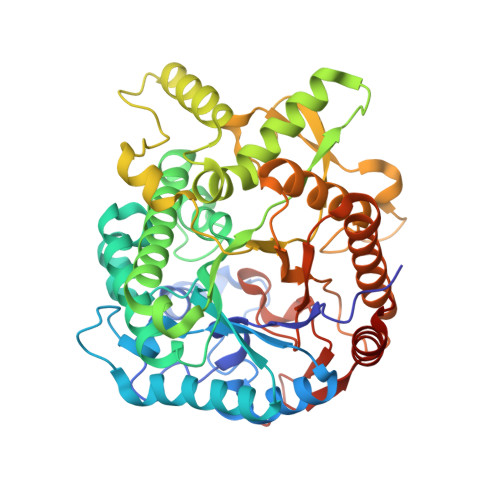
Crystal Structure at 1.1A Resolution of an Insect Myrosinase from Brevicoryne Brassicae Shows its Close Relationship to Beta-Glucosidases.
Husebye, H., Arzt, S., Burmeister, W.P., Haertel, F.V., Brandt, A., Rossiter, J.T., Bones, A.M.(2005) Insect Biochem Mol Biol 35: 1311
- PubMed: 16291087 Search on PubMed
- DOI: https://doi.org/10.1016/j.ibmb.2005.07.004
- Primary Citation Related Structures:
1WCG - PubMed Abstract:
The aphid Brevicoryne brassicae is a specialist feeding on Brassicaceae plants. The insect has an intricate defence system involving a beta-D-thioglucosidase (myrosinase) that hydrolyses glucosinolates sequestered from the host plant into volatile isothiocyanates. These isothiocyanates act synergistically with the pheromone E-beta-farnesene to form an alarm system when the aphid is predated. In order to investigate the enzymatic characteristics of the aphid myrosinase and its three-dimensional structure, milligram amounts of pure recombinant aphid myrosinase were obtained from Echerichia coli. The recombinant enzyme had similar physiochemical properties to the native enzyme. The global structure is very similar to Sinapis alba myrosinase and plant beta-O-glucosidases. Aphid myrosinase has two catalytic glutamic acid residues positioned as in plant beta-O-glucosidases, and it is not obvious why this unusual enzyme hydrolyses glucosinolates, the common substrates of plant myrosinases which are normally not hydrolyzed by plant beta-O-glucosidases. The only residue specific for aphid myrosinase in proximity of the glycosidic linkage is Tyr180 which may have a catalytic role. The aglycon binding site differs strongly from plant myrosinase, whereas due to the presence of Trp424 in the glucose binding site, this part of the active site is more similar to plant beta-O-glucosidases, as plant myrosinases carry a phenylalanine residue at this position.
- Department of Biology, Norwegian University of Science and Technology, Trondheim.
Organizational Affiliation: