1WC8

The crystal structure of mouse bet3p


Experimental Data Snapshot

  • Method: X-RAY DIFFRACTION
  • Resolution: 1.90 Å
  • R-Value Free: 0.260 
  • R-Value Work: 0.217 
  • R-Value Observed: 0.217 

wwPDB Validation   3D Report Full Report


Ligand Structure Quality Assessment 


This is version 1.2 of the entry. See complete history


Literature

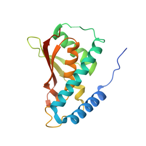
Crystal Structure of Bet3 Reveals a Novel Mechanism for Golgi Localization of Tethering Factor Trapp

Kim, Y.-G.Sohn, E.J.Seo, J.Lee, K.-J.Lee, H.-S.Hwang, I.Whiteway, M.Sacher, M.Oh, B.-H.

(2005) Nat Struct Mol Biol 12: 38

  • DOI: https://doi.org/10.1038/nsmb871
  • Primary Citation of Related Structures:  
    1WC8, 1WC9

  • PubMed Abstract: 

    Transport protein particle (TRAPP) is a large multiprotein complex involved in endoplasmic reticulum-to-Golgi and intra-Golgi traffic. TRAPP specifically and persistently resides on Golgi membranes. Neither the mechanism of the subcellular localization nor the function of any of the individual TRAPP components is known. Here, the crystal structure of mouse Bet3p (bet3), a conserved TRAPP component, reveals a dimeric structure with hydrophobic channels. The channel entrances are located on a putative membrane-interacting surface that is distinctively flat, wide and decorated with positively charged residues. Charge-inversion mutations on the flat surface of the highly conserved yeast Bet3p led to conditional lethality, incorrect localization and membrane trafficking defects. A channel-blocking mutation led to similar defects. These data delineate a molecular mechanism of Golgi-specific targeting and anchoring of Bet3p involving the charged surface and insertion of a Golgi-specific hydrophobic moiety into the channels. This essential subunit could then direct other TRAPP components to the Golgi.


  • Organizational Affiliation

    Center for Biomolecular Recognition, Department of Life Science and Division of Molecular and Life Sciences, Pohang University of Science and Technology, Pohang, Kyungbuk, 790-784, Korea.


Macromolecules
Find similar proteins by:  (by identity cutoff)  |  3D Structure
Entity ID: 1
MoleculeChains Sequence LengthOrganismDetailsImage
TRAFFICKING PROTEIN PARTICLE COMPLEX SUBUNIT3180Mus musculusMutation(s): 0 
UniProt
Find proteins for O55013 (Mus musculus)
Explore O55013 
Go to UniProtKB:  O55013
Entity Groups  
Sequence Clusters30% Identity50% Identity70% Identity90% Identity95% Identity100% Identity
UniProt GroupO55013
Sequence Annotations
Expand
  • Reference Sequence
Small Molecules
Ligands 1 Unique
IDChains Name / Formula / InChI Key2D Diagram3D Interactions
MYR
Query on MYR

Download Ideal Coordinates CCD File 
B [auth A]MYRISTIC ACID
C14 H28 O2
TUNFSRHWOTWDNC-UHFFFAOYSA-N
Experimental Data & Validation

Experimental Data

  • Method: X-RAY DIFFRACTION
  • Resolution: 1.90 Å
  • R-Value Free: 0.260 
  • R-Value Work: 0.217 
  • R-Value Observed: 0.217 
  • Space Group: P 43 21 2
Unit Cell:
Length ( Å )Angle ( ˚ )
a = 54.246α = 90
b = 54.246β = 90
c = 113.757γ = 90
Software Package:
Software NamePurpose
CNSrefinement
SCALEPACKdata scaling

Structure Validation

View Full Validation Report


Ligand Structure Quality Assessment 


Entry History 

Deposition Data

Revision History  (Full details and data files)

  • Version 1.0: 2004-12-13
    Type: Initial release
  • Version 1.1: 2011-05-08
    Changes: Version format compliance
  • Version 1.2: 2011-07-13
    Changes: Version format compliance