1W8L | pdb_00001w8l

Enzymatic and structural characterization of non peptide ligand cyclophilin complexes


Experimental Data Snapshot

  • Method: X-RAY DIFFRACTION
  • Resolution: 1.80 Å
  • R-Value Free: 
    0.230 (Depositor) 
  • R-Value Work: 
    0.179 (Depositor) 
  • R-Value Observed: 
    0.179 (Depositor) 

Starting Model: experimental
View more details

wwPDB Validation 3D Report Full Report

Validation slider image for 1W8L

This is version 1.4 of the entry. See complete history

Literature

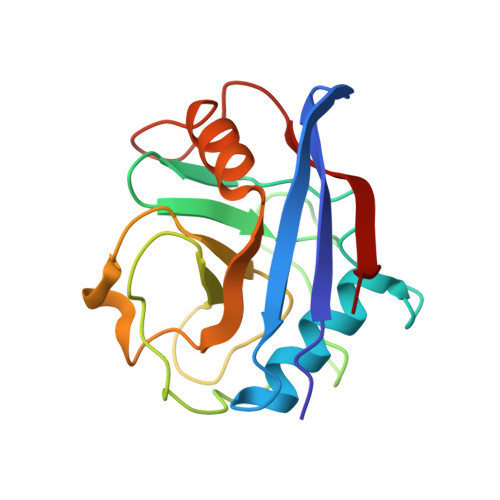
Enzymatic and Structural Characterization of Non-Peptide Ligand-Cyclophilin Complexes

Kontopidis, G.Taylor, P.Walkinshaw, M.

(2004) Acta Crystallogr D Biol Crystallogr 60: 479-485

  • DOI: https://doi.org/10.1107/S0907444904000174
  • Primary Citation Related Structures: 
    1W8L, 1W8M, 1W8V

  • PubMed Abstract: 

    Piperidine ligands are described that provide the first examples of non-peptidic ligand structures for the cyclophilin family of proteins. Crystal structures of two ligand complexes are compared with the unliganded protein and show ligand-induced changes in side-chain conformation and water binding. A peptidylprolyl cis-trans-isomerase assay showed the dissociation constants of the two ligands to be 320 and 25 mM. This study also provides the first published data for both enzymatic activity and three-dimensional structure for any protein-ligand complex that binds with a high-millimolar dissociation constant. The structures may be of relevance in the field of drug design, as they suggest starting points for the design of larger tighter-binding analogues.


  • Organizational Affiliation
    • Structural Biochemistry Group, Department of Biochemistry, The University of Edinburgh, Michael Swann Building, King's Buildings, Edinburgh EH9 3JR, Scotland.

Macromolecule Content 

  • Total Structure Weight: 18.18 kDa 
  • Atom Count: 1,474 
  • Modeled Residue Count: 165 
  • Deposited Residue Count: 165 
  • Unique protein chains: 1

Macromolecules

Find similar proteins by:|  3D Structure
Entity ID: 1
MoleculeChains  Sequence LengthOrganismDetailsImage
PEPTIDYL-PROLYL CIS-TRANS ISOMERASE A165Homo sapiensMutation(s): 0 
EC: 5.2.1.8
UniProt & NIH Common Fund Data Resources
Find proteins for P62937 (Homo sapiens)
Explore P62937 
Go to UniProtKB:  P62937
PHAROS:  P62937
GTEx:  ENSG00000196262 
Entity Groups
Sequence Clusters30% Identity50% Identity70% Identity90% Identity95% Identity100% Identity
UniProt GroupP62937
Sequence Annotations
Expand
Reference Sequence

Small Molecules

Ligands 1 Unique
IDChains Name / Formula / InChI Key2D Diagram3D Interactions
1P3

Query on 1P3


Download:Ideal Coordinates CCD File
B [auth A](3R)-1-ACETYL-3-METHYLPIPERIDINE
C8 H15 N O
XKFPNHDGLSYZRC-SSDOTTSWSA-N

Experimental Data & Validation

Experimental Data

  • Method: X-RAY DIFFRACTION
  • Resolution: 1.80 Å
  • R-Value Free:  0.230 (Depositor) 
  • R-Value Work:  0.179 (Depositor) 
  • R-Value Observed: 0.179 (Depositor) 
Space Group: P 21 21 21
Unit Cell:
Length ( Å )Angle ( ˚ )
a = 36.09α = 90
b = 54.31β = 90
c = 70.97γ = 90
Software Package:
Software NamePurpose
X-PLORrefinement
DENZOdata reduction
SCALEPACKdata scaling
X-PLORphasing

Structure Validation

View Full Validation Report


Entry History 

Deposition Data

Revision History  (Full details and data files)

  • Version 1.0: 2004-09-30
    Type: Initial release
  • Version 1.1: 2011-05-08
    Changes: Version format compliance
  • Version 1.2: 2011-07-13
    Changes: Version format compliance
  • Version 1.3: 2018-06-13
    Changes: Data collection, Database references
  • Version 1.4: 2023-12-13
    Changes: Data collection, Database references, Refinement description