1W17 | pdb_00001w17

Structure of Bacillus subtilis PdaA, a family 4 Carbohydrate esterase.


Experimental Data Snapshot

  • Method: X-RAY DIFFRACTION
  • Resolution: 1.90 Å
  • R-Value Free: 
    0.262 (Depositor), 0.251 (DCC) 
  • R-Value Work: 
    0.201 (Depositor), 0.190 (DCC) 
  • R-Value Observed: 
    0.201 (Depositor) 

wwPDB Validation 3D Report Full Report

Validation slider image for 1W17

This is version 1.5 of the entry. See complete history

Literature

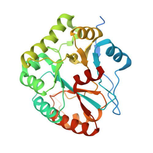
Structures of Bacillus subtilis PdaA, a family 4 carbohydrate esterase, and a complex with N-acetyl-glucosamine.

Blair, D.E.van Aalten, D.M.

(2004) FEBS Lett 570: 13-19

  • DOI: https://doi.org/10.1016/j.febslet.2004.06.013
  • Primary Citation Related Structures: 
    1W17, 1W1A, 1W1B

  • PubMed Abstract: 

    Family 4 carbohydrate esterases deacetylate polymeric carbohydrate substrates such as chitin, acetyl xylan and peptidoglycan. Although some of these enzymes have recently been enzymologically characterised, neither their structure nor their reaction mechanism has been defined. Sequence conservation in this family has pointed to a conserved core, termed the NodB homology domain. We describe the cloning, purification and 1.9 A crystal structure of PdaA, a peptidoglycan deacetylase from Bacillus subtilis. The enzyme assumes a fold related to a (beta/alpha)8 barrel, with a long groove on the surface of the protein that harbours all conserved residues. A complex with the substrate analogue N-acetyl-glucosamine was refined to 2.25 A resolution, revealing interactions of an aspartic acid and three histidines, all conserved in the NodB homology domain, with the ligand. The PdaA structure provides a template for interpreting the wealth of sequence data on family 4 carbohydrate esterases in a structural context and represents a first step towards understanding the reaction mechanism of this family of enzymes.


  • Organizational Affiliation
    • Division of Biological Chemistry and Molecular Microbiology, School of Life Sciences, University of Dundee, Dundee DD1 5EH, UK.

Macromolecule Content 

  • Total Structure Weight: 60.22 kDa 
  • Atom Count: 4,212 
  • Modeled Residue Count: 471 
  • Deposited Residue Count: 526 
  • Unique protein chains: 1

Macromolecules

Find similar proteins by:|  3D Structure
Entity ID: 1
MoleculeChains  Sequence LengthOrganismDetailsImage
PROBABLE POLYSACCHARIDE DEACETYLASE PDAA
A, B
263Bacillus subtilisMutation(s): 0 
EC: 3 (PDB Primary Data), 3.5.1 (UniProt)
UniProt
Find proteins for O34928 (Bacillus subtilis (strain 168))
Explore O34928 
Go to UniProtKB:  O34928
Entity Groups
Sequence Clusters30% Identity50% Identity70% Identity90% Identity95% Identity100% Identity
UniProt GroupO34928
Sequence Annotations
Expand
Reference Sequence

Experimental Data & Validation

Experimental Data

  • Method: X-RAY DIFFRACTION
  • Resolution: 1.90 Å
  • R-Value Free:  0.262 (Depositor), 0.251 (DCC) 
  • R-Value Work:  0.201 (Depositor), 0.190 (DCC) 
  • R-Value Observed: 0.201 (Depositor) 
Space Group: P 65
Unit Cell:
Length ( Å )Angle ( ˚ )
a = 88.115α = 90
b = 88.115β = 90
c = 120.957γ = 120
Software Package:
Software NamePurpose
CNSrefinement
DENZOdata reduction
SCALEPACKdata scaling
MLPHAREphasing

Structure Validation

View Full Validation Report


Entry History 

Deposition Data

Revision History  (Full details and data files)

  • Version 1.0: 2005-01-10
    Type: Initial release
  • Version 1.1: 2011-05-08
    Changes: Version format compliance
  • Version 1.2: 2011-07-13
    Changes: Version format compliance
  • Version 1.3: 2018-02-28
    Changes: Advisory, Database references, Source and taxonomy, Structure summary
  • Version 1.4: 2019-07-24
    Changes: Data collection
  • Version 1.5: 2024-05-08
    Changes: Advisory, Data collection, Database references