1VZG

Structure of superoxide reductase bound to ferrocyanide and active site expansion upon X-ray induced photoreduction


Experimental Data Snapshot

  • Method: X-RAY DIFFRACTION
  • Resolution: 1.69 Å
  • R-Value Free: 0.249 
  • R-Value Work: 0.205 
  • R-Value Observed: 0.205 

wwPDB Validation   3D Report Full Report


This is version 1.3 of the entry. See complete history


Literature

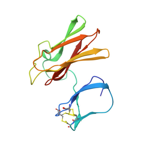
Structure of Superoxide Reductase Bound to Ferrocyanide and Active Site Expansion Upon X-Ray Induced Photoreduction

Adam, V.Royant, A.Niviere, V.Molina-Heredia, F.P.Bourgeois, D.

(2004) Structure 12: 1729

  • DOI: https://doi.org/10.1016/j.str.2004.07.013
  • Primary Citation of Related Structures:  
    1VZG, 1VZH, 1VZI

  • PubMed Abstract: 

    Some sulfate-reducing and microaerophilic bacteria rely on the enzyme superoxide reductase (SOR) to eliminate the toxic superoxide anion radical (O2*-). SOR catalyses the one-electron reduction of O2*- to hydrogen peroxide at a nonheme ferrous iron center. The structures of Desulfoarculus baarsii SOR (mutant E47A) alone and in complex with ferrocyanide were solved to 1.15 and 1.7 A resolution, respectively. The latter structure, the first ever reported of a complex between ferrocyanide and a protein, reveals that this organo-metallic compound entirely plugs the SOR active site, coordinating the active iron through a bent cyano bridge. The subtle structural differences between the mixed-valence and the fully reduced SOR-ferrocyanide adducts were investigated by taking advantage of the photoelectrons induced by X-rays. The results reveal that photo-reduction from Fe(III) to Fe(II) of the iron center, a very rapid process under a powerful synchrotron beam, induces an expansion of the SOR active site.


  • Organizational Affiliation

    LCCP, UMR 5075, IBS-CEA/CNRS/Université J. Fourier, 41 Avenue Jules Horowitz, 38027 Grenoble, Cedex 1, France.


Macromolecules
Find similar proteins by:  (by identity cutoff)  |  3D Structure
Entity ID: 1
MoleculeChains Sequence LengthOrganismDetailsImage
DESULFOFERRODOXIN
A, B
126Desulfarculus baarsiiMutation(s): 1 
EC: 1.15.1.2
UniProt
Find proteins for Q46495 (Desulfarculus baarsii (strain ATCC 33931 / DSM 2075 / LMG 7858 / VKM B-1802 / 2st14))
Explore Q46495 
Go to UniProtKB:  Q46495
Entity Groups  
Sequence Clusters30% Identity50% Identity70% Identity90% Identity95% Identity100% Identity
UniProt GroupQ46495
Sequence Annotations
Expand
  • Reference Sequence
Experimental Data & Validation

Experimental Data

  • Method: X-RAY DIFFRACTION
  • Resolution: 1.69 Å
  • R-Value Free: 0.249 
  • R-Value Work: 0.205 
  • R-Value Observed: 0.205 
  • Space Group: P 21 21 21
Unit Cell:
Length ( Å )Angle ( ˚ )
a = 42.13α = 90
b = 67.65β = 90
c = 82.19γ = 90
Software Package:
Software NamePurpose
CNSrefinement
MOSFLMdata reduction
SCALAdata scaling
CNSphasing

Structure Validation

View Full Validation Report


Entry History 

Deposition Data

Revision History  (Full details and data files)

  • Version 1.0: 2004-08-27
    Type: Initial release
  • Version 1.1: 2011-06-02
    Changes: Version format compliance
  • Version 1.2: 2011-07-13
    Changes: Version format compliance
  • Version 1.3: 2023-12-13
    Changes: Data collection, Database references, Derived calculations, Other, Refinement description