1VZ2

PROLYL OLIGOPEPTIDASE FROM PORCINE BRAIN, Y73C/V427C/C255T MUTANT


Experimental Data Snapshot

  • Method: X-RAY DIFFRACTION
  • Resolution: 2.20 Å
  • R-Value Free: 0.208 
  • R-Value Work: 0.163 
  • R-Value Observed: 0.165 

wwPDB Validation   3D Report Full Report


This is version 1.3 of the entry. See complete history


Literature

Concerted Structural Changes in the Peptidase and the Propeller Domains of Prolyl Oligopeptidase are Required for Substrate Binding

Szeltner, Z.Rea, D.Juhasz, T.Renner, V.Fulop, V.Polgar, L.

(2004) J Mol Biol 340: 627

  • DOI: https://doi.org/10.1016/j.jmb.2004.05.011
  • Primary Citation of Related Structures:  
    1VZ2, 1VZ3

  • PubMed Abstract: 

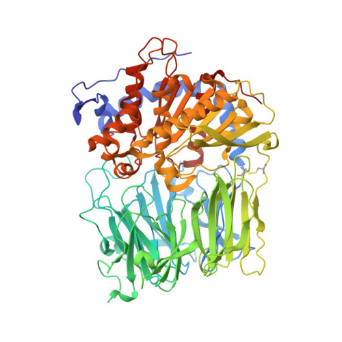
    Prolyl oligopeptidase contains a peptidase domain and its catalytic triad is covered by the central tunnel of a seven-bladed beta-propeller. This domain makes the enzyme an oligopeptidase by excluding large structured peptides from the active site. The apparently rigid crystal structure does not explain how the substrate can approach the catalytic groups. Two possibilities of substrate access were investigated: either blades 1 and 7 of the propeller domain move apart, or the peptidase and/or propeller domains move to create an entry site at the domain interface. Engineering disulfide bridges to the expected oscillating structures prevented such movements, which destroyed the catalytic activity and precluded substrate binding. This indicated that concerted movements of the propeller and the peptidase domains are essential for the enzyme action.


  • Organizational Affiliation

    Institute of Enzymology, Biological Research Center, Hungarian Academy of Sciences, H-1518 Budapest 112, P.O. Box 7, Hungary.


Macromolecules
Find similar proteins by:  (by identity cutoff)  |  3D Structure
Entity ID: 1
MoleculeChains Sequence LengthOrganismDetailsImage
PROLYL ENDOPEPTIDASE710Sus scrofaMutation(s): 3 
EC: 3.4.21.26
UniProt
Find proteins for P23687 (Sus scrofa)
Explore P23687 
Go to UniProtKB:  P23687
Entity Groups  
Sequence Clusters30% Identity50% Identity70% Identity90% Identity95% Identity100% Identity
UniProt GroupP23687
Sequence Annotations
Expand
  • Reference Sequence
Experimental Data & Validation

Experimental Data

  • Method: X-RAY DIFFRACTION
  • Resolution: 2.20 Å
  • R-Value Free: 0.208 
  • R-Value Work: 0.163 
  • R-Value Observed: 0.165 
  • Space Group: P 21 21 21
Unit Cell:
Length ( Å )Angle ( ˚ )
a = 70.9α = 90
b = 99.6β = 90
c = 110γ = 90
Software Package:
Software NamePurpose
REFMACrefinement
DENZOdata reduction
SCALEPACKdata scaling
CCP4phasing

Structure Validation

View Full Validation Report


Entry History 

Deposition Data

Revision History  (Full details and data files)

  • Version 1.0: 2004-07-01
    Type: Initial release
  • Version 1.1: 2011-05-08
    Changes: Version format compliance
  • Version 1.2: 2011-07-13
    Changes: Version format compliance
  • Version 1.3: 2023-12-13
    Changes: Data collection, Database references, Derived calculations, Other, Refinement description