The crystal structure of d(CCCCGGGG): a new A-form variant with an extended backbone conformation.
Haran, T.E., Shakked, Z., Wang, A.H., Rich, A.(1987) J Biomol Struct Dyn 5: 199-217
- PubMed: 3271472 Search on PubMed
- DOI: https://doi.org/10.1080/07391102.1987.10506390
- Primary Citation Related Structures:
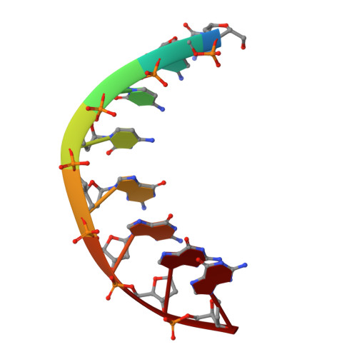
1VT5 - PubMed Abstract:
The crystal structure of d(CCCCGGGG) has been determined at a resolution of 2.25 A. The oligomers crystallize as A-DNA duplexes occupying crystallographic two-fold axes. The backbone conformation is, in general, similar to that observed in previously reported crystal structures of A-DNA fragments, except for the central linkage, where it adopts an extended structure resulting from all trans conformation at the P-O5'-C5'-C4' bonds. This type of conformation facilitates interstrand stacking between the guanines at the C-G site. The local helix twist at this step is very small (25 degrees) compared to an overall average of 33.5 degrees. The unique structure of the C-G base-pair step, namely the extended backbone and the distinct stacking geometry, may be an important feature in the recognition mechanism between double-stranded DNA molecules and restriction endonucleases such as Msp I, which cuts the sequence CCGG very specifically with a rate unaffected by neighboring base pairs.
- Dept. of Structural Chemistry, Weizmann Institute of Science, Rehovot,Israel.
Organizational Affiliation: