1VSR | pdb_00001vsr

VERY SHORT PATCH REPAIR (VSR) ENDONUCLEASE FROM ESCHERICHIA COLI


Experimental Data Snapshot

  • Method: X-RAY DIFFRACTION
  • Resolution: 1.80 Å
  • R-Value Free: 
    0.216 (Depositor), 0.216 (DCC) 
  • R-Value Work: 
    0.196 (Depositor), 0.193 (DCC) 

wwPDB Validation 3D Report Full Report

Validation slider image for 1VSR

This is version 1.3 of the entry. See complete history

Literature

Crystallographic and functional studies of very short patch repair endonuclease.

Tsutakawa, S.E.Muto, T.Kawate, T.Jingami, H.Kunishima, N.Ariyoshi, M.Kohda, D.Nakagawa, M.Morikawa, K.

(1999) Mol Cell 3: 621-628

  • DOI: https://doi.org/10.1016/s1097-2765(00)80355-x
  • Primary Citation Related Structures: 
    1VSR

  • PubMed Abstract: 

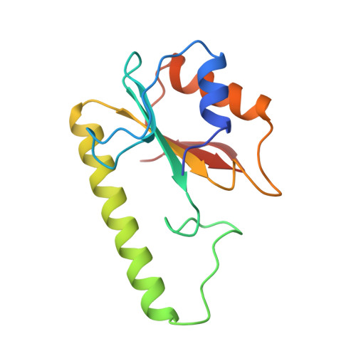
    Vsr endonuclease plays a crucial role in the repair of TG mismatched base pairs, which are generated by the spontaneous degradation of methylated cytidines; Vsr recognizes the mismatched base pair and cleaves the phosphate backbone 5' to the thymidine. We have determined the crystal structure of a truncated form of this endonuclease at 1.8 A resolution. The protein contains one structural zinc-binding module. Unexpectedly, its overall topology resembles members of the type II restriction endonuclease family. Subsequent mutational and biochemical analyses showed that certain elements in the catalytic site are also conserved. However, the identification of a critical histidine and evidence of an active site metal-binding coordination that is novel to endonucleases indicate a distinct catalytic mechanism.


  • Organizational Affiliation
    • Department of Structural Biology, Biomolecular Engineering Research Institute (BERI), Osaka, Japan.

Macromolecule Content 

  • Total Structure Weight: 15.85 kDa 
  • Atom Count: 1,161 
  • Modeled Residue Count: 134 
  • Deposited Residue Count: 136 
  • Unique protein chains: 1

Macromolecules

Find similar proteins by:|  3D Structure
Entity ID: 1
MoleculeChains  Sequence LengthOrganismDetailsImage
PROTEIN (VSR ENDONUCLEASE)136Escherichia coli K-12Mutation(s): 0 
Gene Names: VSR
EC: 3.1
UniProt
Find proteins for P09184 (Escherichia coli (strain K12))
Explore P09184 
Go to UniProtKB:  P09184
Entity Groups
Sequence Clusters30% Identity50% Identity70% Identity90% Identity95% Identity100% Identity
UniProt GroupP09184
Sequence Annotations
Expand
Reference Sequence

Small Molecules

Ligands 1 Unique
IDChains Name / Formula / InChI Key2D Diagram3D Interactions
ZN

Query on ZN


Download:Ideal Coordinates CCD File
B [auth A]ZINC ION
Zn
PTFCDOFLOPIGGS-UHFFFAOYSA-N

Experimental Data & Validation

Experimental Data

  • Method: X-RAY DIFFRACTION
  • Resolution: 1.80 Å
  • R-Value Free:  0.216 (Depositor), 0.216 (DCC) 
  • R-Value Work:  0.196 (Depositor), 0.193 (DCC) 
Space Group: P 21 21 21
Unit Cell:
Length ( Å )Angle ( ˚ )
a = 35.02α = 90
b = 54.24β = 90
c = 73.52γ = 90
Software Package:
Software NamePurpose
CCP4model building
REFMACrefinement
DENZOdata reduction
SCALEPACKdata scaling
CCP4phasing

Structure Validation

View Full Validation Report


Entry History 

Deposition Data

Revision History  (Full details and data files)

  • Version 1.0: 1999-10-27
    Type: Initial release
  • Version 1.1: 2007-10-16
    Changes: Version format compliance
  • Version 1.2: 2011-07-13
    Changes: Source and taxonomy, Version format compliance
  • Version 1.3: 2023-12-27
    Changes: Data collection, Database references, Derived calculations