High-resolution structure of a polyomavirus VP1-oligosaccharide complex: implications for assembly and receptor binding.
Stehle, T., Harrison, S.C.(1997) EMBO J 16: 5139-5148
- PubMed: 9305654 Search on PubMedSearch on PubMed Central
- DOI: https://doi.org/10.1093/emboj/16.16.5139
- Primary Citation Related Structures:
1VPN, 1VPS - PubMed Abstract:
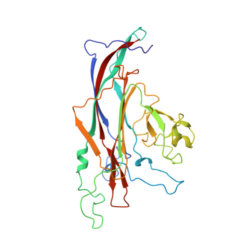
The crystal structure of a recombinant polyomavirus VP1 pentamer (residues 32-320) in complex with a branched disialylated hexasaccharide receptor fragment has been determined at 1.9 A resolution. The result extends our understanding of oligosaccharide receptor recognition. It also suggests a mechanism for enhancing the fidelity of virus assembly. We have previously described the structure of the complete polyomavirus particle complexed with this receptor fragment at 3.65 A. The model presented here offers a much more refined view of the interactions that determine carbohydrate recognition and allows us to assign additional specific contacts, in particular those involving the (alpha2,6)-linked, branching sialic acid. The structure of the unliganded VP1 pentamer, determined independently, shows that the oligosaccharide fits into a preformed groove and induces no measurable structural rearrangements. A comparison with assembled VP1 in the virus capsid reveals a rearrangement of residues 32-45 at the base of the pentamer. This segment may help prevent the formation of incorrectly assembled particles by reducing the likelihood that the C-terminal arm will fold back into its pentamer of origin.
- Howard Hughes Medical Institute and Department of Molecular and Cellular Biology, Harvard University, Cambridge, MA 02138, USA.
Organizational Affiliation: