Mapping the functional surface of insulin by design: structure and function of a novel A-chain analogue.
Hua, Q.X., Hu, S.Q., Frank, B.H., Jia, W., Chu, Y.C., Wang, S.H., Burke, G.T., Katsoyannis, P.G., Weiss, M.A.(1996) J Mol Biology 264: 390-403
- PubMed: 8951384 Search on PubMed
- DOI: https://doi.org/10.1006/jmbi.1996.0648
- Primary Citation Related Structures:
1VKT, 2JMN - PubMed Abstract:
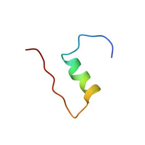
Functional surfaces of a protein are often mapped by combination of X-ray crystallography and mutagenesis. Such studies of insulin have yielded paradoxical results, suggesting that the native state is inactive and reorganizes on receptor binding. Of particular interest is the N-terminal alpha-helix of the A-chain. Does this segment function as an alpha-helix or reorganize as recently proposed in a prohormone-convertase complex? To correlate structure and function, we describe a mapping strategy based on protein design. The solution structure of an engineered monomer ([AspB10, LysB28, ProB29]-human insulin) is determined at neutral pH as a template for synthesis of a novel A-chain analogue. Designed by analogy to a protein-folding intermediate, the analogue lacks the A6-A11 disulphide bridge; the cysteine residues are replaced by serine. Its solution structure is remarkable for segmental unfolding of the N-terminal A-chain alpha-helix (A1 to A8) in an otherwise native subdomain. The structure demonstrates that the overall orientation of the A and B chains is consistent with reorganization of the A-chain's N-terminal segment. Nevertheless, the analogue's low biological activity suggests that this segment, a site of clinical mutation causing diabetes mellitus, functions as a preformed recognition alpha-helix.
- Center for Molecular Oncology, University of Chicago, IL 60637, USA.
Organizational Affiliation: