1VGL | pdb_00001vgl

Crystal structure of tetrameric KaiB from T.elongatus BP-1


Experimental Data Snapshot

  • Method: X-RAY DIFFRACTION
  • Resolution: 2.60 Å
  • R-Value Free: 
    0.289 (Depositor), 0.282 (DCC) 
  • R-Value Work: 
    0.227 (Depositor), 0.223 (DCC) 
  • R-Value Observed: 
    0.230 (Depositor) 

wwPDB Validation 3D Report Full Report

Validation slider image for 1VGL

This is version 1.4 of the entry. See complete history

Literature

Functionally important substructures of circadian clock protein KaiB in a unique tetramer complex.

Iwase, R.Imada, K.Hayashi, F.Uzumaki, T.Morishita, M.Onai, K.Furukawa, Y.Namba, K.Ishiura, M.

(2005) J Biological Chem 280: 43141-43149

  • DOI: https://doi.org/10.1074/jbc.M503360200
  • Primary Citation Related Structures: 
    1VGL

  • PubMed Abstract: 

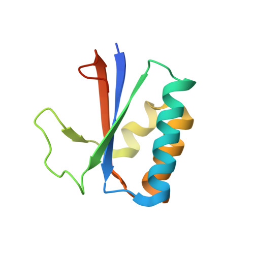
    KaiB is a component of the circadian clock molecular machinery in cyanobacteria, which are the simplest organisms that exhibit circadian rhythms. Here we report the x-ray crystal structure of KaiB from the thermophilic cyanobacterium Thermosynechococcus elongatus BP-1. The KaiB crystal diffracts at a resolution of 2.6 A and includes four subunits organized as a dimer of dimers, each composed of two non-equivalent subunits. The overall shape of the tetramer is an elongated hexagonal plate, with a single positively charged cleft flanked by two negatively charged ridges whose surfaces includes several terminal chains. Site-directed mutagenesis of Synechococcus KaiB confirmed that alanine substitution of residues Lys-11 or Lys-43 in the cleft, or deletion of C-terminal residues 95-108, which forms part of the ridges, strongly weakens in vivo circadian rhythms. Characteristics of KaiB deduced from the x-ray crystal structure were also confirmed by physicochemical measurements of KaiB in solution. These data suggest that the positively charged cleft and flanking negatively charged ridges in KaiB are essential for the biological function of KaiB in the circadian molecular machinery in cyanobacteria.


  • Organizational Affiliation
    • Center for Gene Research, Nagoya University, Furo, Chikusa, Nagoya, Japan.

Macromolecule Content 

  • Total Structure Weight: 48.57 kDa 
  • Atom Count: 3,259 
  • Modeled Residue Count: 404 
  • Deposited Residue Count: 432 
  • Unique protein chains: 1

Macromolecules

Find similar proteins by:|  3D Structure
Entity ID: 1
MoleculeChains  Sequence LengthOrganismDetailsImage
Circadian clock protein kaiB
A, B, C, D
108Thermosynechococcus vestitus BP-1Mutation(s): 1 
UniProt
Find proteins for Q79V61 (Thermosynechococcus vestitus (strain NIES-2133 / IAM M-273 / BP-1))
Explore Q79V61 
Go to UniProtKB:  Q79V61
Entity Groups
Sequence Clusters30% Identity50% Identity70% Identity90% Identity95% Identity100% Identity
UniProt GroupQ79V61
Sequence Annotations
Expand
Reference Sequence

Experimental Data & Validation

Experimental Data

  • Method: X-RAY DIFFRACTION
  • Resolution: 2.60 Å
  • R-Value Free:  0.289 (Depositor), 0.282 (DCC) 
  • R-Value Work:  0.227 (Depositor), 0.223 (DCC) 
  • R-Value Observed: 0.230 (Depositor) 
Space Group: P 1 2 1
Unit Cell:
Length ( Å )Angle ( ˚ )
a = 63.74α = 90
b = 33.41β = 100.12
c = 93.74γ = 90
Software Package:
Software NamePurpose
MOSFLMdata reduction
SCALAdata scaling
SOLVEphasing
REFMACrefinement
CCP4data scaling

Structure Validation

View Full Validation Report


Entry History 

Deposition Data

Revision History  (Full details and data files)

  • Version 1.0: 2005-08-16
    Type: Initial release
  • Version 1.1: 2008-04-27
    Changes: Version format compliance
  • Version 1.2: 2011-07-13
    Changes: Version format compliance
  • Version 1.3: 2021-11-10
    Changes: Database references, Derived calculations
  • Version 1.4: 2023-12-27
    Changes: Data collection