1VDF

ASSEMBLY DOMAIN OF CARTILAGE OLIGOMERIC MATRIX PROTEIN


Experimental Data Snapshot

  • Method: X-RAY DIFFRACTION
  • Resolution: 2.05 Å
  • R-Value Work: 0.176 

wwPDB Validation   3D Report Full Report


This is version 1.2 of the entry. See complete history


Literature

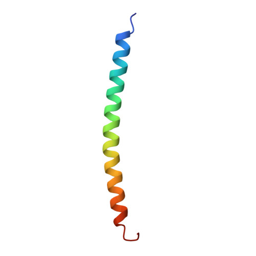
The crystal structure of a five-stranded coiled coil in COMP: a prototype ion channel?

Malashkevich, V.N.Kammerer, R.A.Efimov, V.P.Schulthess, T.Engel, J.

(1996) Science 274: 761-765

  • DOI: 10.1126/science.274.5288.761
  • Primary Citation of Related Structures:  
    1VDF

  • PubMed Abstract: 
  • Oligomerization by the formation of alpha-helical bundles is common in many proteins. The crystal structure of a parallel pentameric coiled coil, constituting the oligomerization domain in the cartilage oligomeric matrix protein (COMP), was determined at 2 ...

    Oligomerization by the formation of alpha-helical bundles is common in many proteins. The crystal structure of a parallel pentameric coiled coil, constituting the oligomerization domain in the cartilage oligomeric matrix protein (COMP), was determined at 2.05 angstroms resolution. The same structure probably occurs in two other extracellular matrix proteins, thrombospondins 3 and 4. Complementary hydrophobic interactions and conserved disulfide bridges between the alpha helices result in a thermostable structure with unusual properties. The long hydrophobic axial pore is filled with water molecules but can also accommodate small apolar groups. An "ion trap" is formed inside the pore by a ring of conserved glutamines, which binds chloride and probably other monatomic anions. The oligomerization domain of COMP has marked similarities with proposed models of the pentameric transmembrane ion channels in phospholamban and the acetylcholine receptor.


    Related Citations: 
    • Crystallization and Preliminary Crystallographic Study of the Pentamerizing Domain from Cartilage Oligomeric Matrix Protein: A Five-Stranded Alpha-Helical Bundle
      Efimov, V.P., Engel, J., Malashkevich, V.N.
      (1996) Proteins 24: 259

    Organizational Affiliation

    Department of Structural Biology, Biozentrum, University of Basel, Klingelbergstrasse 70, CH-4056 Basel, Switzerland.


Macromolecules
Find similar proteins by:  (by identity cutoff)  |  3D Structure
Entity ID: 1
MoleculeChainsSequence LengthOrganismDetailsImage
CARTILAGE OLIGOMERIC MATRIX PROTEINA, B, C, D, E46Rattus norvegicusMutation(s): 0 
Gene Names: Comp
UniProt
Find proteins for P35444 (Rattus norvegicus)
Explore P35444 
Go to UniProtKB:  P35444
Entity Groups  
Sequence Clusters30% Identity50% Identity70% Identity90% Identity95% Identity100% Identity
UniProt GroupP35444
Protein Feature View
Expand
  • Reference Sequence
Small Molecules
Ligands 1 Unique
IDChainsName / Formula / InChI Key2D Diagram3D Interactions
CL
Query on CL

Download Ideal Coordinates CCD File 
F [auth A]CHLORIDE ION
Cl
VEXZGXHMUGYJMC-UHFFFAOYSA-M
 Ligand Interaction
Experimental Data & Validation

Experimental Data

  • Method: X-RAY DIFFRACTION
  • Resolution: 2.05 Å
  • R-Value Work: 0.176 
  • Space Group: P 1 21 1
Unit Cell:
Length ( Å )Angle ( ˚ )
a = 38.47α = 90
b = 49.47β = 103.84
c = 54.98γ = 90
Software Package:
Software NamePurpose
MOSFLMdata reduction
SCALAdata scaling
MLPHAREphasing
TNTrefinement
CCP4data scaling

Structure Validation

View Full Validation Report


Entry History 

Deposition Data

Revision History  (Full details and data files)

  • Version 1.0: 1997-10-08
    Type: Initial release
  • Version 1.1: 2008-03-25
    Changes: Version format compliance
  • Version 1.2: 2011-07-13
    Changes: Version format compliance