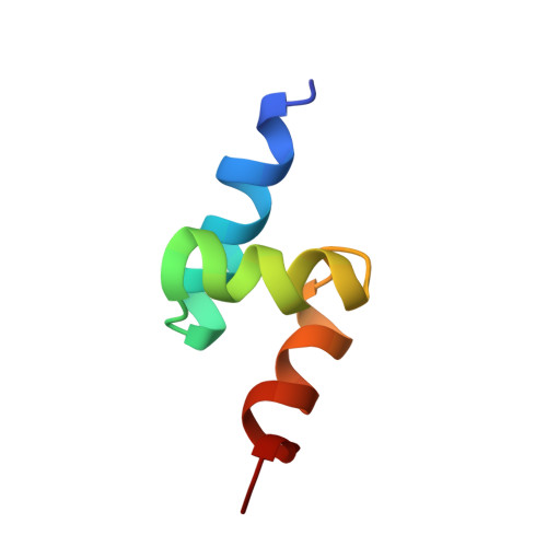
1V92 | pdb_00001v92

Solution structure of the UBA domain from p47, a major cofactor of the AAA ATPase p97


Experimental Data Snapshot

  • Method: SOLUTION NMR
  • Conformers Calculated: 100 
  • Conformers Submitted: 10 
  • Selection Criteria: structures with acceptable covalent geometry,structures with favorable non-bond energy,structures with the least restraint violations,structures with the lowest energy 

wwPDB Validation 3D Report Full Report

Validation slider image for 1V92

This is version 1.4 of the entry. See complete history

Literature

Structure, dynamics and interactions of p47, a major adaptor of the AAA ATPase, p97

Yuan, X.Simpson, P.McKeown, C.Kondo, H.Uchiyama, K.Wallis, R.Dreveny, I.Keetch, C.Zhang, X.Robinson, C.Freemont, P.Matthews, S.

(2004) EMBO J 23: 1463-1473

  • DOI: https://doi.org/10.1038/sj.emboj.7600152
  • Primary Citation Related Structures: 
    1V92, 1VAZ

  • PubMed Abstract: 

    p47 is a major adaptor molecule of the cytosolic AAA ATPase p97. The principal role of the p97-p47 complex is in regulation of membrane fusion events. Mono-ubiquitin recognition by p47 has also been shown to be crucial in the p97-p47-mediated Golgi membrane fusion events. Here, we describe the high-resolution solution structures of the N-terminal UBA domain and the central domain (SEP) from p47. The p47 UBA domain has the characteristic three-helix bundle fold and forms a highly stable complex with ubiquitin. We report the interaction surfaces of the two proteins and present a structure for the p47 UBA-ubiquitin complex. The p47 SEP domain adopts a novel fold with a betabetabetaalphaalphabeta secondary structure arrangement, where beta4 pairs in a parallel fashion to beta1. Based on biophysical studies, we demonstrate a clear propensity for the self-association of p47. Furthermore, p97 N binding abolishes p47 self-association, revealing the potential interaction surfaces for recognition of other domains within p97 or the substrate.


  • Organizational Affiliation
    • Department of Biological Sciences, Wolfson Laboratories, Imperial College London, South Kensington, London, UK.

Macromolecule Content 

  • Total Structure Weight: 5.17 kDa 
  • Atom Count: 365 
  • Modeled Residue Count: 46 
  • Deposited Residue Count: 46 
  • Unique protein chains: 1

Macromolecules

Find similar proteins by:|  3D Structure
Entity ID: 1
MoleculeChains  Sequence LengthOrganismDetailsImage
NSFL1 cofactor p4746Rattus norvegicusMutation(s): 0 
UniProt
Find proteins for O35987 (Rattus norvegicus)
Explore O35987 
Go to UniProtKB:  O35987
Entity Groups
Sequence Clusters30% Identity50% Identity70% Identity90% Identity95% Identity100% Identity
UniProt GroupO35987
Sequence Annotations
Expand
Reference Sequence

Experimental Data & Validation

Experimental Data

  • Method: SOLUTION NMR
  • Conformers Calculated: 100 
  • Conformers Submitted: 10 
  • Selection Criteria: structures with acceptable covalent geometry,structures with favorable non-bond energy,structures with the least restraint violations,structures with the lowest energy 

Structure Validation

View Full Validation Report


Entry History 

Deposition Data

Revision History  (Full details and data files)

  • Version 1.0: 2004-04-06
    Type: Initial release
  • Version 1.1: 2008-04-27
    Changes: Version format compliance
  • Version 1.2: 2011-07-13
    Changes: Version format compliance
  • Version 1.3: 2022-03-02
    Changes: Data collection, Database references, Derived calculations
  • Version 1.4: 2023-12-27
    Changes: Data collection