1V8X

Crystal Structure of the Dioxygen-bound Heme Oxygenase from Corynebacterium diphtheriae


Experimental Data Snapshot

  • Method: X-RAY DIFFRACTION
  • Resolution: 1.85 Å
  • R-Value Free: 0.193 
  • R-Value Work: 0.153 
  • R-Value Observed: 0.157 

wwPDB Validation   3D Report Full Report


Ligand Structure Quality Assessment 


This is version 2.1 of the entry. See complete history


Literature

Crystal Structure of the Dioxygen-bound Heme Oxygenase from Corynebacterium diphtheriae: IMPLICATIONS FOR HEME OXYGENASE FUNCTION.

Unno, M.Matsui, T.Chu, G.C.Couture, M.Yoshida, T.Rousseau, D.L.Olson, J.S.Ikeda-Saito, M.

(2004) J Biol Chem 279: 21055-21061

  • DOI: https://doi.org/10.1074/jbc.M400491200
  • Primary Citation of Related Structures:  
    1V8X

  • PubMed Abstract: 

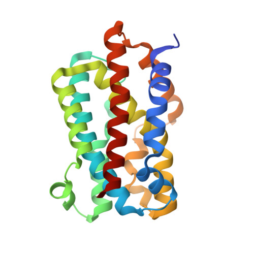
    HmuO, a heme oxygenase of Corynebacterium diphtheriae, catalyzes degradation of heme using the same mechanism as the mammalian enzyme. The oxy form of HmuO, the precursor of the catalytically active ferric hydroperoxo species, has been characterized by ligand binding kinetics, resonance Raman spectroscopy, and x-ray crystallography. The oxygen association and dissociation rate constants are 5 microm(-1) s(-1) and 0.22 s(-1), respectively, yielding an O(2) affinity of 21 microm(-1), which is approximately 20 times greater than that of mammalian myoglobins. However, the affinity of HmuO for CO is only 3-4-fold greater than that for mammalian myoglobins, implying the presence of strong hydrogen bonding interactions in the distal pocket of HmuO that preferentially favor O(2) binding. Resonance Raman spectra show that the Fe-O(2) vibrations are tightly coupled to porphyrin vibrations, indicating the highly bent Fe-O-O geometry that is characteristic of the oxy forms of heme oxygenases. In the crystal structure of the oxy form the Fe-O-O angle is 110 degrees, the O-O bond is pointed toward the heme alpha-meso-carbon by direct steric interactions with Gly-135 and Gly-139, and hydrogen bonds occur between the bound O(2) and the amide nitrogen of Gly-139 and a distal pocket water molecule, which is a part of an extended hydrogen bonding network that provides the solvent protons required for oxygen activation. In addition, the O-O bond is orthogonal to the plane of the proximal imidazole side chain, which facilitates hydroxylation of the porphyrin alpha-meso-carbon by preventing premature O-O bond cleavage.


  • Organizational Affiliation

    Institute of Multidisciplinary Research for Advanced Materials, Tohoku University, Katahira, Aoba, Sendai 980-8577, Japan. unno19@tagen.tohoku.ac.jp


Macromolecules
Find similar proteins by:  (by identity cutoff)  |  3D Structure
Entity ID: 1
MoleculeChains Sequence LengthOrganismDetailsImage
Heme oxygenase
A, B, C
215Corynebacterium diphtheriaeMutation(s): 0 
EC: 1.14.99.3
UniProt
Find proteins for P71119 (Corynebacterium diphtheriae (strain ATCC 700971 / NCTC 13129 / Biotype gravis))
Explore P71119 
Go to UniProtKB:  P71119
Entity Groups  
Sequence Clusters30% Identity50% Identity70% Identity90% Identity95% Identity100% Identity
UniProt GroupP71119
Sequence Annotations
Expand
  • Reference Sequence
Oligosaccharides

Help

Entity ID: 2
MoleculeChains Length2D Diagram Glycosylation3D Interactions
beta-D-fructofuranose-(2-1)-alpha-D-glucopyranose
D
2N/A
Glycosylation Resources
GlyTouCan:  G05551OP
GlyCosmos:  G05551OP
Small Molecules
Ligands 3 Unique
IDChains Name / Formula / InChI Key2D Diagram3D Interactions
HEM
Query on HEM

Download Ideal Coordinates CCD File 
E [auth A],
J [auth B],
P [auth C]
PROTOPORPHYRIN IX CONTAINING FE
C34 H32 Fe N4 O4
KABFMIBPWCXCRK-RGGAHWMASA-L
SO4
Query on SO4

Download Ideal Coordinates CCD File 
F [auth A]
G [auth A]
H [auth A]
K [auth B]
L [auth B]
F [auth A],
G [auth A],
H [auth A],
K [auth B],
L [auth B],
M [auth B],
N [auth B],
Q [auth C],
R [auth C],
S [auth C],
T [auth C],
U [auth C]
SULFATE ION
O4 S
QAOWNCQODCNURD-UHFFFAOYSA-L
OXY
Query on OXY

Download Ideal Coordinates CCD File 
I [auth A],
O [auth B],
V [auth C]
OXYGEN MOLECULE
O2
MYMOFIZGZYHOMD-UHFFFAOYSA-N
Biologically Interesting Molecules (External Reference) 1 Unique
Experimental Data & Validation

Experimental Data

  • Method: X-RAY DIFFRACTION
  • Resolution: 1.85 Å
  • R-Value Free: 0.193 
  • R-Value Work: 0.153 
  • R-Value Observed: 0.157 
  • Space Group: P 1 21 1
Unit Cell:
Length ( Å )Angle ( ˚ )
a = 54.003α = 90
b = 62.969β = 100.96
c = 107.49γ = 90
Software Package:
Software NamePurpose
REFMACrefinement
SCALEPACKdata scaling
CNSphasing

Structure Validation

View Full Validation Report


Ligand Structure Quality Assessment 


Entry History 

Deposition Data

Revision History  (Full details and data files)

  • Version 1.0: 2004-05-18
    Type: Initial release
  • Version 1.1: 2008-04-27
    Changes: Version format compliance
  • Version 1.2: 2011-07-13
    Changes: Version format compliance
  • Version 1.3: 2017-10-04
    Changes: Refinement description
  • Version 2.0: 2020-07-29
    Type: Remediation
    Reason: Carbohydrate remediation
    Changes: Atomic model, Data collection, Derived calculations, Non-polymer description, Structure summary
  • Version 2.1: 2023-12-27
    Changes: Data collection, Database references, Structure summary