1V6M

Peanut Lectin with 9mer peptide (IWSSAGNVA)


Experimental Data Snapshot

  • Method: X-RAY DIFFRACTION
  • Resolution: 2.70 Å
  • R-Value Free: 0.264 
  • R-Value Work: 0.210 
  • R-Value Observed: 0.210 

wwPDB Validation   3D Report Full Report


This is version 1.3 of the entry. See complete history


Literature

Structural plasticity of peanut lectin: an X-ray analysis involving variation in pH, ligand binding and crystal structure.

Kundhavai Natchiar, S.Arockia Jeyaprakash, A.Ramya, T.N.Thomas, C.J.Suguna, K.Surolia, A.Vijayan, M.

(2004) Acta Crystallogr D Biol Crystallogr 60: 211-219

  • DOI: https://doi.org/10.1107/S090744490302849X
  • Primary Citation of Related Structures:  
    1V6I, 1V6J, 1V6K, 1V6L, 1V6M, 1V6N, 1V6O

  • PubMed Abstract: 

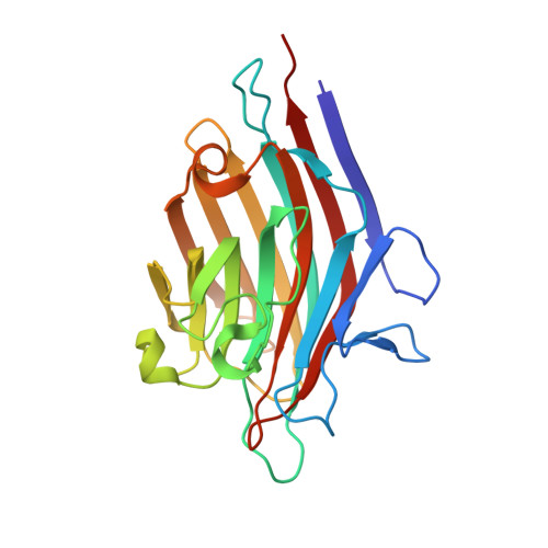
    Until recently, it has only been possible to grow crystals of peanut lectin when complexed with sugar ligands. It is now shown that it is possible to grow peanut lectin crystals at acidic pH in the presence of oligopeptides corresponding to a loop in the lectin molecule. Crystals have also been prepared in the presence of these peptides as well as lactose. Low-pH crystal forms of the lectin-lactose complex similar to those obtained at neutral pH have also been grown. Thus, crystals of peanut lectin grown under different environmental conditions, at two pH values with and without sugar bound to the lectin, are now available. They have been used to explore the plasticity and hydration of the molecule. A detailed comparison between different structures shows that the lectin molecule is sturdy and that the effect of changes in pH, ligand binding and environment on it is small. The region involving the curved front beta-sheet and the loops around the second hydrophobic core is comparatively rigid. The back beta-sheet involved in quaternary association, which exhibits considerable variability, is substantially flexible, as is the sugar-binding region. The numbers of invariant water molecules in the hydration shell are small and they are mainly involved in metal coordination or in stabilizing unusual structural features. Small consistent movements occur in the combining site upon sugar binding, although the site is essentially preformed.


  • Organizational Affiliation

    Molecular Biophysics Unit, Indian Institute of Science, Bangalore 560 012, India.


Macromolecules
Find similar proteins by:  (by identity cutoff)  |  3D Structure
Entity ID: 1
MoleculeChains Sequence LengthOrganismDetailsImage
Galactose-binding lectin
A, B, C, D, E
A, B, C, D, E, F, G, H
232Arachis hypogaeaMutation(s): 0 
UniProt
Find proteins for P02872 (Arachis hypogaea)
Explore P02872 
Go to UniProtKB:  P02872
Entity Groups  
Sequence Clusters30% Identity50% Identity70% Identity90% Identity95% Identity100% Identity
UniProt GroupP02872
Sequence Annotations
Expand
  • Reference Sequence
Small Molecules
Ligands 2 Unique
IDChains Name / Formula / InChI Key2D Diagram3D Interactions
MN
Query on MN

Download Ideal Coordinates CCD File 
J [auth A]
L [auth B]
N [auth C]
P [auth D]
R [auth E]
J [auth A],
L [auth B],
N [auth C],
P [auth D],
R [auth E],
T [auth F],
V [auth G],
X [auth H]
MANGANESE (II) ION
Mn
WAEMQWOKJMHJLA-UHFFFAOYSA-N
CA
Query on CA

Download Ideal Coordinates CCD File 
I [auth A]
K [auth B]
M [auth C]
O [auth D]
Q [auth E]
I [auth A],
K [auth B],
M [auth C],
O [auth D],
Q [auth E],
S [auth F],
U [auth G],
W [auth H]
CALCIUM ION
Ca
BHPQYMZQTOCNFJ-UHFFFAOYSA-N
Experimental Data & Validation

Experimental Data

  • Method: X-RAY DIFFRACTION
  • Resolution: 2.70 Å
  • R-Value Free: 0.264 
  • R-Value Work: 0.210 
  • R-Value Observed: 0.210 
  • Space Group: P 1 21 1
Unit Cell:
Length ( Å )Angle ( ˚ )
a = 127.852α = 90
b = 126.139β = 116.16
c = 84.825γ = 90
Software Package:
Software NamePurpose
CNSrefinement
DENZOdata reduction
SCALEPACKdata scaling

Structure Validation

View Full Validation Report


Entry History 

Deposition Data

Revision History  (Full details and data files)

  • Version 1.0: 2004-02-10
    Type: Initial release
  • Version 1.1: 2008-04-27
    Changes: Version format compliance
  • Version 1.2: 2011-07-13
    Changes: Version format compliance
  • Version 1.3: 2023-10-25
    Changes: Data collection, Database references, Derived calculations, Refinement description