1V6B | pdb_00001v6b

Solution structure of the third PDZ domain of mouse harmonin


Experimental Data Snapshot

  • Method: SOLUTION NMR
  • Conformers Calculated: 100 
  • Conformers Submitted: 20 
  • Selection Criteria: target function 

wwPDB Validation 3D Report Full Report

Validation slider image for 1V6B

This is version 1.4 of the entry. See complete history

Literature

Solution structure of the third PDZ domain of mouse harmonin

Yamada, K.Nameki, N.Saito, K.Koshiba, S.Inoue, M.Kigawa, T.Yokoyama, S.

To be published.

Macromolecule Content 

  • Total Structure Weight: 12.46 kDa 
  • Atom Count: 875 
  • Modeled Residue Count: 118 
  • Deposited Residue Count: 118 
  • Unique protein chains: 1

Macromolecules

Find similar proteins by:|  3D Structure
Entity ID: 1
MoleculeChains  Sequence LengthOrganismDetailsImage
harmonin isoform a1118Mus musculusMutation(s): 0 
Gene Names: RIKEN cDNA 2010016F01
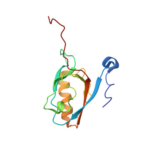
UniProt & NIH Common Fund Data Resources
Find proteins for Q9ES64 (Mus musculus)
Explore Q9ES64 
Go to UniProtKB:  Q9ES64
IMPC:  MGI:1919338
Entity Groups
Sequence Clusters30% Identity50% Identity70% Identity90% Identity95% Identity100% Identity
UniProt GroupQ9ES64
Sequence Annotations
Expand
Reference Sequence

Experimental Data & Validation

Experimental Data

  • Method: SOLUTION NMR
  • Conformers Calculated: 100 
  • Conformers Submitted: 20 
  • Selection Criteria: target function 

Structure Validation

View Full Validation Report


Entry History 

Revision History  (Full details and data files)

  • Version 1.0: 2004-05-28
    Type: Initial release
  • Version 1.1: 2008-04-27
    Changes: Version format compliance
  • Version 1.2: 2011-07-13
    Changes: Version format compliance
  • Version 1.3: 2022-03-02
    Changes: Data collection, Database references, Derived calculations
  • Version 1.4: 2023-12-27
    Changes: Data collection