1V4U

Crystal structure of bluefin tuna carbonmonoxy-hemoglobin


Experimental Data Snapshot

  • Method: X-RAY DIFFRACTION
  • Resolution: 2.00 Å
  • R-Value Free: 0.264 
  • R-Value Work: 0.204 
  • R-Value Observed: 0.207 

wwPDB Validation   3D Report Full Report


This is version 1.3 of the entry. See complete history


Literature

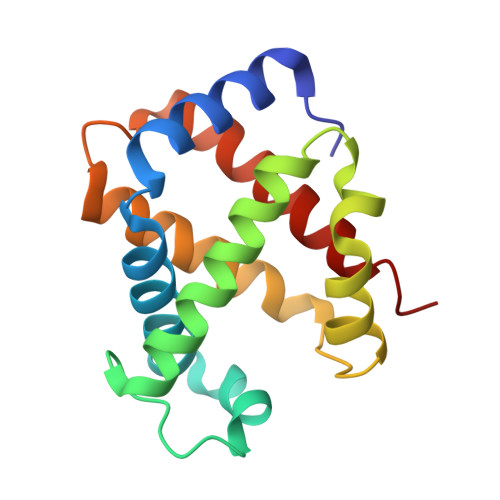
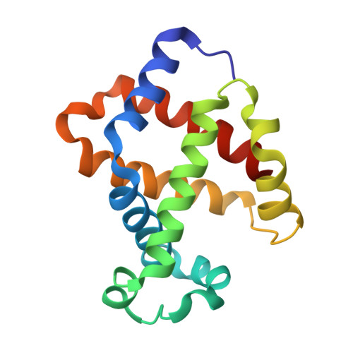
Novel Mechanisms of pH Sensitivity in Tuna Hemoglobin: A STRUCTURAL EXPLANATION OF THE ROOT EFFECT

Yokoyama, T.Chong, K.T.Miyazaki, G.Morimoto, H.Shih, D.T.Unzai, S.Tame, J.R.Park, S.Y.

(2004) J Biol Chem 279: 28632-28640

  • DOI: https://doi.org/10.1074/jbc.M401740200
  • Primary Citation of Related Structures:  
    1V4U, 1V4W, 1V4X

  • PubMed Abstract: 

    The crystal structure of hemoglobin has been known for several decades, yet various features of the molecule remain unexplained or controversial. Several animal hemoglobins have properties that cannot be readily explained in terms of their amino acid sequence and known atomic models of hemoglobin. Among these, fish hemoglobins are well known for their widely varying interactions with heterotropic effector molecules and pH sensitivity. Some fish hemoglobins are almost completely insensitive to pH (within physiological limits), whereas others show extremely low oxygen affinity under acid conditions, a phenomenon called the Root effect. X-ray crystal structures of Root effect hemoglobins have not, to date, provided convincing explanations of this effect. Sequence alignments have signally failed to pinpoint the residues involved, and site-directed mutagenesis has not yielded a human hemoglobin variant with this property. We have solved the crystal structure of tuna hemoglobin in the deoxy form at low and moderate pH and in the presence of carbon monoxide at high pH. A comparison of these models shows clear evidence for novel mechanisms of pH-dependent control of ligand affinity.


  • Organizational Affiliation

    Protein Design Laboratory, Yokohama City University, Suehiro-cho 1-7-29, Tsurumi, Yokohama 230-0045, Japan.


Macromolecules
Find similar proteins by:  (by identity cutoff)  |  3D Structure
Entity ID: 1
MoleculeChains Sequence LengthOrganismDetailsImage
hemoglobin alpha chain
A, C
144Thunnus thynnusMutation(s): 0 
UniProt
Find proteins for Q8AYM0 (Thunnus thynnus)
Explore Q8AYM0 
Go to UniProtKB:  Q8AYM0
Entity Groups  
Sequence Clusters30% Identity50% Identity70% Identity90% Identity95% Identity100% Identity
UniProt GroupQ8AYM0
Sequence Annotations
Expand
  • Reference Sequence
Find similar proteins by:  (by identity cutoff)  |  3D Structure
Entity ID: 2
MoleculeChains Sequence LengthOrganismDetailsImage
hemoglobin beta chain
B, D
146Thunnus thynnusMutation(s): 0 
UniProt
Find proteins for Q8AYM1 (Thunnus thynnus)
Explore Q8AYM1 
Go to UniProtKB:  Q8AYM1
Entity Groups  
Sequence Clusters30% Identity50% Identity70% Identity90% Identity95% Identity100% Identity
UniProt GroupQ8AYM1
Sequence Annotations
Expand
  • Reference Sequence
Experimental Data & Validation

Experimental Data

  • Method: X-RAY DIFFRACTION
  • Resolution: 2.00 Å
  • R-Value Free: 0.264 
  • R-Value Work: 0.204 
  • R-Value Observed: 0.207 
  • Space Group: P 21 21 21
Unit Cell:
Length ( Å )Angle ( ˚ )
a = 59.016α = 90
b = 102.541β = 90
c = 108.451γ = 90
Software Package:
Software NamePurpose
REFMACrefinement
HKL-2000data reduction
SCALEPACKdata scaling
MOLREPphasing

Structure Validation

View Full Validation Report


Entry History 

Deposition Data

Revision History  (Full details and data files)

  • Version 1.0: 2004-07-06
    Type: Initial release
  • Version 1.1: 2008-04-27
    Changes: Version format compliance
  • Version 1.2: 2011-07-13
    Changes: Version format compliance
  • Version 1.3: 2023-12-27
    Changes: Data collection, Database references, Derived calculations