1V3M

Crystal structure of F283Y mutant cyclodextrin glycosyltransferase complexed with a pseudo-tetraose derived from acarbose


Experimental Data Snapshot

  • Method: X-RAY DIFFRACTION
  • Resolution: 2.00 Å
  • R-Value Free: 0.218 
  • R-Value Work: 0.163 
  • R-Value Observed: 0.167 

wwPDB Validation   3D Report Full Report


Ligand Structure Quality Assessment 


This is version 2.2 of the entry. See complete history


Literature

Role of Phe283 in enzymatic reaction of cyclodextrin glycosyltransferase from alkalophilic Bacillus sp.1011: Substrate binding and arrangement of the catalytic site

Kanai, R.Haga, K.Akiba, T.Yamane, K.Harata, K.

(2004) Protein Sci 13: 457-465

  • DOI: https://doi.org/10.1110/ps.03408504
  • Primary Citation of Related Structures:  
    1V3J, 1V3K, 1V3L, 1V3M

  • PubMed Abstract: 

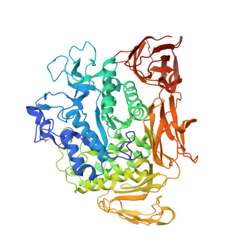
    Cyclodextrin glycosyltransferase (CGTase) belonging to the alpha-amylase family mainly catalyzes transglycosylation and produces cyclodextrins from starch and related alpha-1,4-glucans. The catalytic site of CGTase specifically conserves four aromatic residues, Phe183, Tyr195, Phe259, and Phe283, which are not found in alpha-amylase. To elucidate the structural role of Phe283, we determined the crystal structures of native and acarbose-complexed mutant CGTases in which Phe283 was replaced with leucine (F283L) or tyrosine (F283Y). The temperature factors of the region 259-269 in native F283L increased >10 A(2) compared with the wild type. The complex formation with acarbose not only increased the temperature factors (>10 A(2)) but also changed the structure of the region 257-267. This region is stabilized by interactions of Phe283 with Phe259 and Leu260 and plays an important role in the cyclodextrin binding. The conformation of the side-chains of Glu257, Phe259, His327, and Asp328 in the catalytic site was altered by the mutation of Phe283 with leucine, and this indicates that Phe283 partly arranges the structure of the catalytic site through contacts with Glu257 and Phe259. The replacement of Phe283 with tyrosine decreased the enzymatic activity in the basic pH range. The hydroxyl group of Tyr283 forms hydrogen bonds with the carboxyl group of Glu257, and the pK(a) of Glu257 in F283Y may be lower than that in the wild type.


  • Organizational Affiliation

    Biological Information Research Center, AIST Tsukuba Central 6, 1-1-1 Higashi, Tsukuba, Ibaraki 305-8566, Japan.


Macromolecules
Find similar proteins by:  (by identity cutoff)  |  3D Structure
Entity ID: 1
MoleculeChains Sequence LengthOrganismDetailsImage
Cyclomaltodextrin glucanotransferase
A, B
686Bacillus sp. 1011Mutation(s): 1 
EC: 2.4.1.19
UniProt
Find proteins for P05618 (Bacillus sp. (strain 1011))
Explore P05618 
Go to UniProtKB:  P05618
Entity Groups  
Sequence Clusters30% Identity50% Identity70% Identity90% Identity95% Identity100% Identity
UniProt GroupP05618
Sequence Annotations
Expand
  • Reference Sequence
Oligosaccharides

Help

Entity ID: 2
MoleculeChains Length2D Diagram Glycosylation3D Interactions
4,6-dideoxy-alpha-D-xylo-hexopyranose-(1-4)-beta-D-galactopyranose
C, D
2N/A
Glycosylation Resources
GlyTouCan:  G94251DX
GlyCosmos:  G94251DX
Entity ID: 3
MoleculeChains Length2D Diagram Glycosylation3D Interactions
alpha-D-glucopyranose-(1-4)-alpha-D-glucopyranose
E
2N/A
Glycosylation Resources
GlyTouCan:  G07411ON
GlyCosmos:  G07411ON
Small Molecules
Ligands 4 Unique
IDChains Name / Formula / InChI Key2D Diagram3D Interactions
GAL
Query on GAL

Download Ideal Coordinates CCD File 
J [auth A],
O [auth B],
P [auth B]
beta-D-galactopyranose
C6 H12 O6
WQZGKKKJIJFFOK-FPRJBGLDSA-N
GLC
Query on GLC

Download Ideal Coordinates CCD File 
F [auth A],
H [auth A],
I [auth A],
M [auth B]
alpha-D-glucopyranose
C6 H12 O6
WQZGKKKJIJFFOK-DVKNGEFBSA-N
ACI
Query on ACI

Download Ideal Coordinates CCD File 
G [auth A],
N [auth B]
6-AMINO-4-HYDROXYMETHYL-CYCLOHEX-4-ENE-1,2,3-TRIOL
C7 H13 N O4
XPHOBMULWMGEBA-VZFHVOOUSA-N
CA
Query on CA

Download Ideal Coordinates CCD File 
K [auth A],
L [auth A],
Q [auth B],
R [auth B]
CALCIUM ION
Ca
BHPQYMZQTOCNFJ-UHFFFAOYSA-N
Biologically Interesting Molecules (External Reference) 1 Unique
Experimental Data & Validation

Experimental Data

  • Method: X-RAY DIFFRACTION
  • Resolution: 2.00 Å
  • R-Value Free: 0.218 
  • R-Value Work: 0.163 
  • R-Value Observed: 0.167 
  • Space Group: P 1
Unit Cell:
Length ( Å )Angle ( ˚ )
a = 64.09α = 84.93
b = 73.6β = 105.12
c = 78.3γ = 101
Software Package:
Software NamePurpose
FFFEARdata reduction
X-PLORmodel building
X-PLORrefinement
SMARTdata reduction
FFFEARdata scaling
X-PLORphasing

Structure Validation

View Full Validation Report


Ligand Structure Quality Assessment 


Entry History 

Deposition Data

Revision History  (Full details and data files)

  • Version 1.0: 2004-08-03
    Type: Initial release
  • Version 1.1: 2007-10-16
    Changes: Version format compliance
  • Version 1.2: 2011-07-13
    Changes: Source and taxonomy, Version format compliance
  • Version 1.3: 2017-10-04
    Changes: Refinement description
  • Version 2.0: 2020-07-29
    Type: Remediation
    Reason: Carbohydrate remediation
    Changes: Advisory, Atomic model, Data collection, Derived calculations, Structure summary
  • Version 2.1: 2021-11-10
    Changes: Database references, Structure summary
  • Version 2.2: 2023-10-25
    Changes: Data collection, Refinement description