1V18

The crystal structure of beta-catenin armadillo repeat complexed with a phosphorylated APC 20mer repeat.


Experimental Data Snapshot

  • Method: X-RAY DIFFRACTION
  • Resolution: 2.10 Å
  • R-Value Free: 0.253 
  • R-Value Work: 0.207 
  • R-Value Observed: 0.207 

wwPDB Validation   3D Report Full Report


This is version 1.3 of the entry. See complete history


Literature

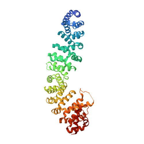
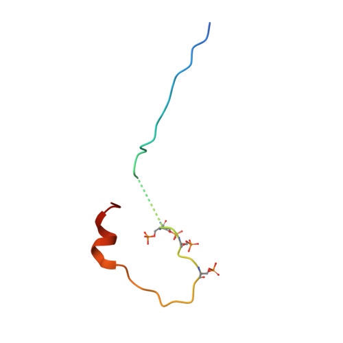
Mechanism of Phosphorylation-Dependent Binding of Apc to Beta-Catenin and its Role in Beta-Catenin Degradation

Ha, N.-C.Tonozuka, T.Stamos, J.L.Weis, W.I.

(2004) Mol Cell 15: 511

  • DOI: https://doi.org/10.1016/j.molcel.2004.08.010
  • Primary Citation of Related Structures:  
    1T08, 1V18

  • PubMed Abstract: 

    The transcriptional coactivator beta-catenin mediates Wnt growth factor signaling. In the absence of a Wnt signal, casein kinase 1 (CK1) and glycogen synthase kinase-3beta (GSK-3beta) phosphorylate cytosolic beta-catenin, thereby flagging it for recognition and destruction by the ubiquitin/proteosome machinery. Phosphorylation occurs in a multiprotein complex that includes the kinases, beta-catenin, axin, and the Adenomatous Polyposis Coli (APC) protein. The role of APC in this process is poorly understood. CK1epsilon and GSK-3beta phosphorylate APC, which increases its affinity for beta-catenin. Crystal structures of phosphorylated and nonphosphorylated APC bound to beta-catenin reveal a phosphorylation-dependent binding motif generated by mutual priming of CK1 and GSK-3beta substrate sequences. Axin is shown to act as a scaffold for substrate phosphorylation by these kinases. Phosphorylated APC and axin bind to the same surface of, and compete directly for, beta-catenin. The structural and biochemical data suggest a novel model for how APC functions in beta-catenin degradation.


  • Organizational Affiliation

    Department of Structural Biology, Stanford University School of Medicine, Stanford, CA 94043, USA.


Macromolecules
Find similar proteins by:  (by identity cutoff)  |  3D Structure
Entity ID: 1
MoleculeChains Sequence LengthOrganismDetailsImage
BETA-CATENIN538Mus musculusMutation(s): 0 
UniProt & NIH Common Fund Data Resources
Find proteins for Q02248 (Mus musculus)
Explore Q02248 
Go to UniProtKB:  Q02248
IMPC:  MGI:88276
Entity Groups  
Sequence Clusters30% Identity50% Identity70% Identity90% Identity95% Identity100% Identity
UniProt GroupQ02248
Sequence Annotations
Expand
  • Reference Sequence
Find similar proteins by:  (by identity cutoff)  |  3D Structure
Entity ID: 2
MoleculeChains Sequence LengthOrganismDetailsImage
ADENOMATOUS POLYPOSIS COLI47Homo sapiensMutation(s): 0 
UniProt & NIH Common Fund Data Resources
Find proteins for P25054 (Homo sapiens)
Explore P25054 
Go to UniProtKB:  P25054
PHAROS:  P25054
GTEx:  ENSG00000134982 
Entity Groups  
Sequence Clusters30% Identity50% Identity70% Identity90% Identity95% Identity100% Identity
UniProt GroupP25054
Sequence Annotations
Expand
  • Reference Sequence
Small Molecules
Modified Residues  1 Unique
IDChains TypeFormula2D DiagramParent
SEP
Query on SEP
B
L-PEPTIDE LINKINGC3 H8 N O6 PSER
Experimental Data & Validation

Experimental Data

  • Method: X-RAY DIFFRACTION
  • Resolution: 2.10 Å
  • R-Value Free: 0.253 
  • R-Value Work: 0.207 
  • R-Value Observed: 0.207 
  • Space Group: P 21 21 21
Unit Cell:
Length ( Å )Angle ( ˚ )
a = 50.83α = 90
b = 90.06β = 90
c = 122.81γ = 90
Software Package:
Software NamePurpose
CNSrefinement
HKL-2000data reduction
HKL-2000data scaling
AMoREphasing

Structure Validation

View Full Validation Report


Entry History 

Deposition Data

Revision History  (Full details and data files)

  • Version 1.0: 2005-01-12
    Type: Initial release
  • Version 1.1: 2011-05-08
    Changes: Version format compliance
  • Version 1.2: 2011-07-13
    Changes: Version format compliance
  • Version 1.3: 2023-12-13
    Changes: Data collection, Database references, Derived calculations, Other, Refinement description