1V05

Dimerization of human Filamin C: crystal structure of the domain 24


Experimental Data Snapshot

  • Method: X-RAY DIFFRACTION
  • Resolution: 1.43 Å
  • R-Value Free: 0.205 
  • R-Value Work: 0.186 
  • R-Value Observed: 0.188 

wwPDB Validation   3D Report Full Report


This is version 1.3 of the entry. See complete history


Literature

Structural Basis for Vertebrate Filamin Dimerization

Pudas, R.Kiema, T.-R.Butler, P.J.G.Stewart, M.Ylanne, J.

(2005) Structure 13: 111

  • DOI: https://doi.org/10.1016/j.str.2004.10.014
  • Primary Citation of Related Structures:  
    1V05

  • PubMed Abstract: 

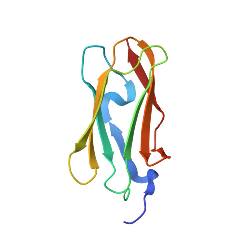
    Filamins are essential in cell motility and many developmental processes. They are large actin cross linking proteins that contain actin binding domains in their N termini and a long rod region constructed from 24 tandem Ig domains. Dimerization is crucial for the actin crosslinking function of filamins and requires the most C-terminal Ig domain. We describe here the crystal structure of this 24th Ig domain (Ig24) of human filamin C and show how it mediates dimerization. The dimer interface is novel and quite different to that seen in the Dictyostelium discoideum filamin analog. The sequence signature of the dimerization interface suggests that the C-terminal domains of all vertebrate filamins share the same dimerization mechanism. Furthermore, we show that point mutations in the dimerization interface disrupt the dimer and that the dissociation constant for recombinant Ig24 is in the micromolar range.


  • Organizational Affiliation

    Biocenter Oulu and Department of Biochemistry, University of Oulu, Finland.


Macromolecules
Find similar proteins by:  (by identity cutoff)  |  3D Structure
Entity ID: 1
MoleculeChains Sequence LengthOrganismDetailsImage
FILAMIN C96Homo sapiensMutation(s): 0 
UniProt & NIH Common Fund Data Resources
Find proteins for Q14315 (Homo sapiens)
Explore Q14315 
Go to UniProtKB:  Q14315
PHAROS:  Q14315
GTEx:  ENSG00000128591 
Entity Groups  
Sequence Clusters30% Identity50% Identity70% Identity90% Identity95% Identity100% Identity
UniProt GroupQ14315
Sequence Annotations
Expand
  • Reference Sequence
Experimental Data & Validation

Experimental Data

  • Method: X-RAY DIFFRACTION
  • Resolution: 1.43 Å
  • R-Value Free: 0.205 
  • R-Value Work: 0.186 
  • R-Value Observed: 0.188 
  • Space Group: P 61 2 2
Unit Cell:
Length ( Å )Angle ( ˚ )
a = 48.099α = 90
b = 48.099β = 90
c = 117.485γ = 120
Software Package:
Software NamePurpose
REFMACrefinement
XDSdata reduction
XDSdata scaling
CNSphasing

Structure Validation

View Full Validation Report


Entry History 

Deposition Data

Revision History  (Full details and data files)

  • Version 1.0: 2004-11-17
    Type: Initial release
  • Version 1.1: 2011-05-07
    Changes: Version format compliance
  • Version 1.2: 2011-07-13
    Changes: Version format compliance
  • Version 1.3: 2019-07-24
    Changes: Data collection