1UW5 | pdb_00001uw5

Structure of PITP-alpha complexed to phosphatidylinositol


Experimental Data Snapshot

  • Method: X-RAY DIFFRACTION
  • Resolution: 2.90 Å
  • R-Value Free: 
    0.307 (Depositor) 
  • R-Value Work: 
    0.237 (Depositor) 

Starting Model: experimental
View more details

wwPDB Validation 3D Report Full Report

Validation slider image for 1UW5

This is version 1.5 of the entry. See complete history

Literature

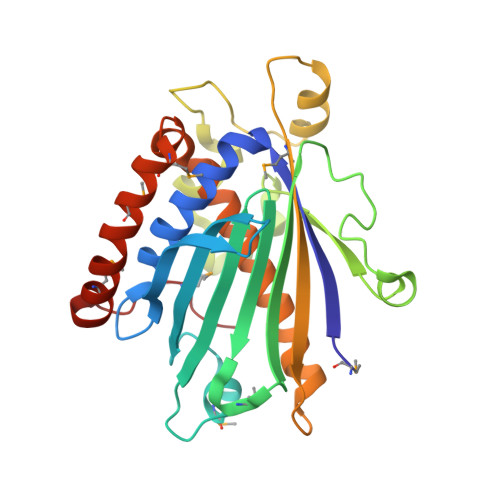
Structure-Function Analysis of Human [Corrected] Phosphatidylinositol Transfer Protein Alpha Bound to Phosphatidylinositol.

Tilley, S.J.Skippen, A.Murray-Rust, J.Swigart, P.M.Stewart, A.Morgan, C.P.Cockcroft, S.Mcdonald, N.Q.

(2004) Structure 12: 317

  • DOI: https://doi.org/10.1016/j.str.2004.01.013
  • Primary Citation Related Structures: 
    1UW5

  • PubMed Abstract: 

    Phosphatidylinositol transfer protein alpha (PITPalpha) selectively transports and promotes exchange of phosphatidylinositol (PI) and phosphatidylcholine (PC) between lipid bilayers. In higher eukaryotes PITPalpha is required for cellular functions such as phospholipase C-mediated signaling, regulated exocytosis, and secretory vesicle formation. We have determined the crystal structure of human PITPalpha bound to its physiological ligand, PI, at 2.95 A resolution. The structure identifies the critical side chains within the lipid-headgroup binding pocket that define the exquisite specificity for PI. Mutational analysis of the PI binding pocket is in good agreement with the structural data and allows manipulation of functional properties of PITPalpha. Surprisingly, there are no major conformational differences between PI- and PC-loaded PITPalpha, despite previous predictions. In the crystal, PITPalpha-PI is dimeric, with two identical dimers in the asymmetric unit. The dimer interface masks precisely the sequence we identify as contributing to PITPalpha membrane interaction. Our structure represents a soluble, transport-competent form of PI-loaded PITPalpha.


  • Organizational Affiliation
    • School of Crystallography, Birkbeck, University of London, Malet Street, London WC1E 7HX, UK.

Macromolecule Content 

  • Total Structure Weight: 132.26 kDa 
  • Atom Count: 8,727 
  • Modeled Residue Count: 1,075 
  • Deposited Residue Count: 1,080 
  • Unique protein chains: 1

Macromolecules

Find similar proteins by:|  3D Structure
Entity ID: 1
MoleculeChains  Sequence LengthOrganismDetailsImage
PHOSPHATIDYLINOSITOL TRANSFER PROTEIN ALPHA ISOFORM
A, B, C, D
270Homo sapiensMutation(s): 0 
UniProt & NIH Common Fund Data Resources
Find proteins for Q00169 (Homo sapiens)
Explore Q00169 
Go to UniProtKB:  Q00169
PHAROS:  Q00169
GTEx:  ENSG00000174238 
Entity Groups
Sequence Clusters30% Identity50% Identity70% Identity90% Identity95% Identity100% Identity
UniProt GroupQ00169
Sequence Annotations
Expand
Reference Sequence

Small Molecules

Modified Residues  1 Unique
IDChains TypeFormula2D DiagramParent
MSE
Query on MSE
A, B, C, D
L-PEPTIDE LINKINGC5 H11 N O2 SeMET

Experimental Data & Validation

Experimental Data

  • Method: X-RAY DIFFRACTION
  • Resolution: 2.90 Å
  • R-Value Free:  0.307 (Depositor) 
  • R-Value Work:  0.237 (Depositor) 
Space Group: P 21 21 21
Unit Cell:
Length ( Å )Angle ( ˚ )
a = 83.262α = 90
b = 93.173β = 90
c = 161.732γ = 90
Software Package:
Software NamePurpose
REFMACrefinement
MOSFLMdata reduction
SCALAdata scaling
MOLREPphasing

Structure Validation

View Full Validation Report


Entry History 

Deposition Data

Revision History  (Full details and data files)

  • Version 1.0: 2004-03-04
    Type: Initial release
  • Version 1.1: 2011-05-08
    Changes: Version format compliance
  • Version 1.2: 2011-07-13
    Changes: Version format compliance
  • Version 1.3: 2023-12-13
    Changes: Data collection, Database references, Derived calculations, Refinement description
  • Version 1.4: 2024-10-23
    Changes: Structure summary
  • Version 1.5: 2025-12-24
    Changes: Derived calculations, Structure summary