1UW0 | pdb_00001uw0

Solution structure of the zinc-finger domain from DNA ligase IIIa


Experimental Data Snapshot

  • Method: SOLUTION NMR
  • Conformers Calculated: 50 
  • Conformers Submitted: 28 
  • Selection Criteria: LOW RESTRAINT VIOLATIONS 

wwPDB Validation 3D Report Full Report

Validation slider image for 1UW0

This is version 1.3 of the entry. See complete history

Literature

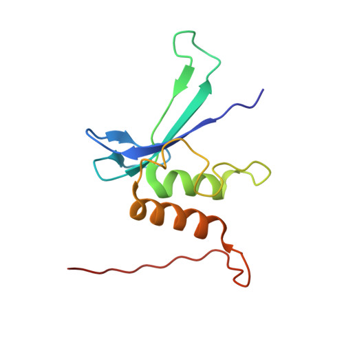
Solution Structure and DNA Binding of the Zinc-Finger Domain from DNA Ligase Iiialpha

Kulczyk, A.W.Yang, J.-C.Neuhaus, D.

(2004) J Mol Biology 341: 723

  • DOI: https://doi.org/10.1016/j.jmb.2004.06.035
  • Primary Citation Related Structures: 
    1UW0

  • PubMed Abstract: 

    DNA ligase IIIalpha carries out the final ligation step in the base excision repair (BER) and single strand break repair (SSBR) mechanisms of DNA repair. The enzyme recognises single-strand nicks and other damage features in double-stranded DNA, both through the catalytic domain and an N-terminal domain containing a single zinc finger. The latter is homologous to other zinc fingers that recognise damaged DNA, two in the N terminus of poly(adenosine-ribose)polymerase and three in the N terminus of the Arabidopsis thaliana nick-sensing DNA 3'-phosphoesterase. Here, we present the solution structure of the zinc-finger domain of human DNA ligase IIIalpha, the first structure of a finger from this group. It is related to that of the erythroid transcription factor GATA-1, but has an additional N-terminal beta-strand and C-terminal alpha-helix. Chemical shift mapping using a DNA ligand containing a single-stranded break showed that the DNA-binding surface of the DNA-ligase IIIalpha zinc finger is substantially different from that of GATA-1, consistent with the fact that the two proteins recognise very different features in the DNA. Likely implications for DNA binding are discussed.


  • Organizational Affiliation
    • MRC Laboratory of Molecular Biology, Hills Road, Cambridge CB2 2QH, UK.

Macromolecule Content 

  • Total Structure Weight: 13.31 kDa 
  • Atom Count: 924 
  • Modeled Residue Count: 117 
  • Deposited Residue Count: 117 
  • Unique protein chains: 1

Macromolecules

Find similar proteins by:|  3D Structure
Entity ID: 1
MoleculeChains  Sequence LengthOrganismDetailsImage
DNA LIGASE III117Homo sapiensMutation(s): 0 
EC: 6.5.1.1
UniProt & NIH Common Fund Data Resources
Find proteins for P49916 (Homo sapiens)
Explore P49916 
Go to UniProtKB:  P49916
PHAROS:  P49916
GTEx:  ENSG00000005156 
Entity Groups
Sequence Clusters30% Identity50% Identity70% Identity90% Identity95% Identity100% Identity
UniProt GroupP49916
Sequence Annotations
Expand
Reference Sequence

Small Molecules

Ligands 1 Unique
IDChains Name / Formula / InChI Key2D Diagram3D Interactions
ZN

Query on ZN


Download:Ideal Coordinates CCD File
B [auth A]ZINC ION
Zn
PTFCDOFLOPIGGS-UHFFFAOYSA-N

Experimental Data & Validation

Experimental Data

  • Method: SOLUTION NMR
  • Conformers Calculated: 50 
  • Conformers Submitted: 28 
  • Selection Criteria: LOW RESTRAINT VIOLATIONS 

Structure Validation

View Full Validation Report


Entry History 

Deposition Data

Revision History  (Full details and data files)

  • Version 1.0: 2004-08-05
    Type: Initial release
  • Version 1.1: 2011-05-08
    Changes: Version format compliance
  • Version 1.2: 2011-07-13
    Changes: Version format compliance
  • Version 1.3: 2024-05-15
    Changes: Data collection, Database references, Derived calculations, Other