1UUE | pdb_00001uue

a-SPECTRIN SH3 DOMAIN (V44T, D48G MUTANT)


Experimental Data Snapshot

  • Method: X-RAY DIFFRACTION
  • Resolution: 2.60 Å
  • R-Value Free: 
    0.266 (Depositor) 
  • R-Value Work: 
    0.240 (Depositor), 0.250 (DCC) 
  • R-Value Observed: 
    0.240 (Depositor) 

Starting Model: experimental
View more details

wwPDB Validation 3D Report Full Report

Validation slider image for 1UUE

This is version 1.7 of the entry. See complete history

Literature

Solvation in Protein Folding Analysis: Combination of Theoretical and Experimental Approaches

Fernandez, A.Vega, M.C.Wilmanns, M.Serrano, L.

(2004) Proc Natl Acad Sci U S A 101: 2834

  • DOI: https://doi.org/10.1073/pnas.0304180101
  • Primary Citation Related Structures: 
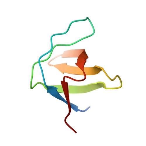
    1UUE

  • PubMed Abstract: 

    An effort to combine theoretical analyses and protein engineering methods has been made to probe the folding mechanism of SH3 by using Energy Landscape Theory and a phi-value analysis. Particular emphasis was given to core residues and the effect of desolvation during the folding event by replacing the core valines with isosteric threonines. These mutations have the advantage of keeping the core structurally invariant while affecting core stability relative to the unfolded state. Although the valines that form the core appear spatially invariant, the folding kinetics of their threonine mutants varies, indicating their different extent of solvation in the transition-state ensemble. Theoretical studies predicted the distribution of folding kinetics of threonine mutants without previous knowledge of the measured rates. This initial success encourages further investigations of the molecular details behind these macroscopic phenomena and of the role of solvation in the folding mechanism.


  • Organizational Affiliation
    • European Molecular Biology Laboratory, Meyerhofstrasse 1, D-69117, Heidelberg, Germany.

Macromolecule Content 

  • Total Structure Weight: 7.17 kDa 
  • Atom Count: 492 
  • Modeled Residue Count: 57 
  • Deposited Residue Count: 62 
  • Unique protein chains: 1

Macromolecules

Find similar proteins by:|  3D Structure
Entity ID: 1
MoleculeChains  Sequence LengthOrganismDetailsImage
SPECTRIN ALPHA CHAIN62Gallus gallusMutation(s): 2 
UniProt
Find proteins for P07751 (Gallus gallus)
Explore P07751 
Go to UniProtKB:  P07751
Entity Groups
Sequence Clusters30% Identity50% Identity70% Identity90% Identity95% Identity100% Identity
UniProt GroupP07751
Sequence Annotations
Expand
Reference Sequence

Experimental Data & Validation

Experimental Data

  • Method: X-RAY DIFFRACTION
  • Resolution: 2.60 Å
  • R-Value Free:  0.266 (Depositor) 
  • R-Value Work:  0.240 (Depositor), 0.250 (DCC) 
  • R-Value Observed: 0.240 (Depositor) 
Space Group: P 21 21 21
Unit Cell:
Length ( Å )Angle ( ˚ )
a = 33.094α = 90
b = 41.877β = 90
c = 49.616γ = 90
Software Package:
Software NamePurpose
CNSrefinement
MOSFLMdata reduction
SCALAdata scaling
AMoREphasing

Structure Validation

View Full Validation Report


Entry History 

Deposition Data

Revision History  (Full details and data files)

  • Version 1.0: 2004-02-19
    Type: Initial release
  • Version 1.1: 2011-05-08
    Changes: Version format compliance
  • Version 1.2: 2011-07-13
    Changes: Version format compliance
  • Version 1.3: 2018-10-24
    Changes: Data collection, Source and taxonomy
  • Version 1.4: 2019-07-10
    Changes: Data collection
  • Version 1.5: 2019-07-24
    Changes: Data collection
  • Version 1.6: 2019-10-16
    Changes: Data collection, Experimental preparation, Other
  • Version 1.7: 2023-12-13
    Changes: Data collection, Database references, Refinement description